Skip to content
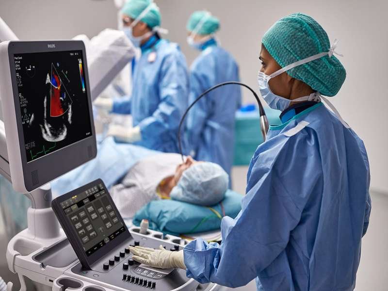
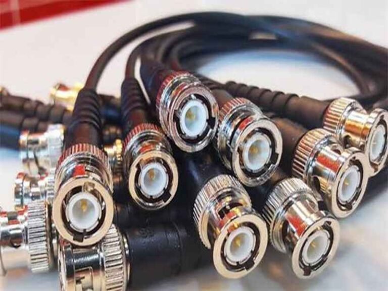
Ultrasound equipment depends on very weak, high-frequency signals. These signals travel from the transducer probe to the imaging system through a cable assembly that must remain stable while the probe is moved, rotated, cleaned, pulled, and used every day. If the cable structure is not correct, the equipment may still power on, but image quality can suffer. Noise, signal loss, poor shielding, unstable impedance, cable fatigue, or connector wear can all affect the final diagnostic image.
Cable solutions for ultrasound equipment include probe cables, micro coax cable assemblies, shielded signal cables, RF cables, LVDS cables, power cables, and internal harnesses. These cables are designed to transmit high-frequency signals with low loss, reduce EMI interference, support repeated movement, fit compact device structures, and maintain long-term reliability in medical environments.
For ultrasound OEMs, cable selection is not only about finding a connector and cable length. It involves frequency, impedance, shielding, OD, flexibility, bending life, connector availability, material compliance, and production consistency. A cable that works in a first prototype may still fail after months of use if the strain relief, shielding, or conductor structure is not designed for the real working environment.
Sino-Conn often receives ultrasound cable inquiries where customers only provide a sample, connector photo, or partial drawing. In these cases, our first step is not to quote blindly. We review the cable structure, signal requirement, connector direction, shielding method, and mechanical stress points before preparing drawings and samples. This is especially important for ultrasound projects, because small cable design details can directly affect imaging stability and user experience.
What Are Ultrasound Cables?
Ultrasound cables are specialized cable assemblies designed to transmit electrical signals, power, control commands, and imaging data within ultrasound systems. They connect probes, transducers, beamformers, imaging processors, displays, power modules, and internal electronic boards.

Unlike ordinary electronic cables, ultrasound cables work with extremely weak and sensitive signals. Even small changes in impedance, shielding effectiveness, conductor quality, or connector performance can influence the final image displayed on the screen.
This is why ultrasound cable assemblies are considered critical components rather than simple accessories.
For an ultrasound manufacturer, the transducer, electronics, software, and cable assembly form a complete signal chain. The overall imaging performance is often limited by the weakest part of that chain.
Probe Cables
Probe cables are the most recognizable ultrasound cables because they are directly connected to the transducer used by doctors and sonographers.

Every time an examination is performed, the probe cable moves continuously.
A single hospital ultrasound system may perform:
| Usage Level | Estimated Scans Per Year |
|---|---|
| Small Clinic | 2,000–5,000 |
| Regional Hospital | 5,000–15,000 |
| Large Medical Center | 15,000–30,000+ |
During these examinations, the cable experiences:
- Continuous bending
- Twisting
- Pulling
- Rotation
- Compression
- Cleaning cycles
Over several years, the cable may experience hundreds of thousands of flex cycles.
This explains why probe cable design focuses heavily on:
- Flexibility
- Fatigue resistance
- Signal stability
- Strain relief design
- User comfort
Many people assume ultrasound probe failures are caused by the transducer itself.
In reality, service centers frequently find that cable-related damage is responsible for a significant percentage of probe repairs.
Common failure locations include:
| Failure Area | Typical Cause |
|---|---|
| Probe Exit | Repeated bending |
| Connector End | Pulling stress |
| Internal Conductors | Fatigue damage |
| Shield Layer | Long-term flexing |
| Strain Relief | Mechanical wear |
One customer sent Sino-Conn several failed probe cable samples from a portable ultrasound platform.
At first glance, all samples appeared normal externally.
After analysis, multiple conductors were found to have fractured internally near the strain relief area.
The root cause was not the conductor itself.
The issue was excessive stress concentration caused by the original cable geometry.
After modifying the strain relief structure and conductor design, cable lifespan increased substantially during validation testing.
This illustrates an important reality:
A probe cable is both an electrical component and a mechanical component.
Success depends on both.
Micro Coax
Micro coax cable technology has become one of the most important developments in modern ultrasound systems.

As imaging quality improves, probe manufacturers continue increasing channel counts.
Higher channel counts allow:
- Better image resolution
- Improved focusing
- Enhanced contrast
- More advanced beamforming
However, more channels also create a packaging problem.
The cable cannot become excessively thick or heavy.
Doctors need lightweight probes that remain comfortable during long scanning sessions.
This is where micro coax becomes essential.
Each micro coax line contains:
- Center conductor
- Dielectric insulation
- Individual shield
- Outer insulation
Because every channel is individually shielded, signal isolation improves significantly.
| Cable Structure | Crosstalk Control |
|---|---|
| Multi-core Wire | Moderate |
| Shielded Twisted Pair | Good |
| Micro Coax | Excellent |
Modern premium ultrasound probes may contain:
| Probe Category | Approximate Signal Channels |
|---|---|
| Basic Probe | 64–128 |
| General Imaging Probe | 128–192 |
| Advanced Probe | 192–256 |
| High-End Systems | 256–512+ |
Without micro coax technology, these channel counts would be extremely difficult to package into a flexible cable assembly.
One ultrasound OEM approached Sino-Conn while developing a handheld scanner.
The engineering team wanted to reduce overall probe weight while maintaining imaging performance.
After evaluating multiple cable structures, a micro coax solution allowed channel density to increase while reducing overall cable diameter.
The final design improved both ergonomics and signal performance.
Signal Cables
Signal transmission is the primary purpose of most ultrasound cable assemblies.
The signals generated inside the transducer are extremely small.
Protecting these signals during transmission is one of the biggest engineering challenges in ultrasound equipment design.
Several factors directly affect signal quality:
- Cable impedance
- Conductor resistance
- Shield coverage
- Dielectric stability
- Connector quality
- Assembly consistency
Even a minor variation can influence performance.
| Parameter | Why It Matters |
|---|---|
| Impedance | Controls reflections |
| Attenuation | Preserves signal strength |
| Shielding | Reduces noise |
| Conductor Quality | Improves transmission |
| Connector Stability | Maintains consistency |
Many customers focus heavily on transducer specifications.
However, a high-performance transducer connected to a poor-quality signal cable may never achieve its intended performance.
A useful comparison is to think of the cable as a highway.
Even if the vehicle is excellent, traffic flow suffers when the road is poorly designed.
Signal cables must be designed to carry information efficiently from the probe to the processing electronics without introducing unnecessary losses.
Power Cables
Power cables are sometimes overlooked because they do not directly contribute to image generation.
However, stable power delivery is essential for reliable system operation.
Modern ultrasound systems contain:
- High-speed processors
- Digital imaging boards
- Displays
- Cooling systems
- Battery modules
- Data communication systems
All require stable power.
Poor power cable design may contribute to:
- Voltage drops
- Heat generation
- Startup issues
- Reduced reliability
| Design Factor | Impact |
|---|---|
| Wire Gauge | Current capacity |
| Connector Resistance | Heat generation |
| Cable Length | Voltage drop |
| Insulation Quality | Safety |
| Flexibility | Installation |
Portable ultrasound systems create additional challenges because designers want:
- Smaller cables
- Lower weight
- Higher charging capability
- Longer battery life
These goals often compete with one another.
Finding the right balance requires careful cable engineering rather than simply increasing conductor size.
Internal Harnesses
Many people think only about the cable attached to the probe.
In reality, modern ultrasound systems contain multiple cable assemblies inside the equipment.
A typical ultrasound platform may include:
- Probe interface harnesses
- Display cables
- Power harnesses
- LVDS assemblies
- RF cables
- Communication cables
- Battery connections
These internal harnesses connect dozens of electronic modules.
As ultrasound systems become more compact, internal cable routing becomes increasingly complex.
Engineers must balance:
- Space constraints
- EMI performance
- Serviceability
- Manufacturing efficiency
- Long-term reliability
| Internal Cable Type | Common Function |
|---|---|
| LVDS Cable | Display transmission |
| Micro Coax | High-frequency signals |
| RF Cable | Signal routing |
| Power Harness | Power distribution |
| USB Assembly | Service interfaces |
| FFC/FPC | Compact board connections |
One medical equipment manufacturer approached Sino-Conn because assembly times were increasing on a new ultrasound platform.
The original internal harness design required technicians to route several cable branches through tight spaces during production.
After reviewing the mechanical layout, branch locations and connector orientations were redesigned.
The result was faster assembly, improved service access, and lower installation error rates.
This highlights a point that is often overlooked:
Ultrasound cables do much more than transmit signals.
They influence manufacturing efficiency, maintenance costs, product reliability, user comfort, and ultimately the overall success of the ultrasound system itself.
The best ultrasound cable assemblies are designed not only for electrical performance but also for how the equipment will be assembled, used, cleaned, serviced, and operated over many years in clinical environments.
Why Do Ultrasound Cables Matter?
For many people outside the medical device industry, an ultrasound cable appears to be a simple connection between the probe and the imaging system.
For ultrasound engineers, however, the cable assembly is one of the most critical components in the entire signal path.
Every image displayed on the monitor begins as an electrical signal generated inside the transducer. Before software processing, beamforming, image enhancement, or AI-assisted analysis can occur, those signals must first travel through the cable assembly.
If signal quality is compromised during transmission, the imaging system can never fully recover the lost information.
This is why leading ultrasound manufacturers invest significant engineering resources into cable design, shielding, connector systems, and material selection.
A well-designed ultrasound cable contributes to:
- Higher image quality
- Better signal consistency
- Lower EMI susceptibility
- Improved probe handling
- Longer service life
- Reduced maintenance costs
- Better user experience
A poorly designed cable may lead to:
- Image artifacts
- Signal instability
- Probe failures
- Increased warranty claims
- More service repairs
- Customer complaints
In many cases, cable-related issues do not appear during initial testing. They emerge after months or years of clinical use, making them even more expensive to address.
Image Quality
Image quality is ultimately the reason ultrasound systems exist.
Doctors rely on image clarity to identify anatomical structures, detect abnormalities, guide procedures, and make clinical decisions.
The cable assembly plays a direct role in preserving the integrity of the signal before it reaches the imaging electronics.
Several cable-related factors influence image quality:
| Parameter | Impact on Imaging |
|---|---|
| Signal Loss | Reduced image detail |
| Crosstalk | Blurred boundaries |
| Noise Pickup | Increased artifacts |
| Impedance Variation | Signal reflection |
| Shielding Gaps | Image instability |
The challenge becomes more significant as imaging systems become more advanced.
Modern ultrasound systems often support:
- Higher frequencies
- More imaging channels
- Better resolution
- Faster processing
As performance increases, signal margins become smaller.
A cable structure that worked adequately ten years ago may no longer meet the requirements of modern imaging platforms.
One ultrasound manufacturer contacted Sino-Conn after noticing inconsistent image quality between production units.
The transducers, PCBs, and software were identical.
After investigation, slight variations in cable assembly construction were identified as the source of the problem.
By standardizing cable materials, conductor structures, and shielding termination methods, image consistency improved across the entire production batch.
This illustrates an important point:
Image quality is not determined solely by the transducer.
The cable connecting that transducer to the imaging system is equally important.
Signal Loss
Signal loss is one of the most common engineering challenges in ultrasound cable design.
The signals generated by transducer elements are often extremely small.
Any loss occurring between the probe and the imaging processor reduces the amount of usable information available for image reconstruction.
Several factors contribute to signal loss:
- Conductor resistance
- Cable length
- Frequency
- Dielectric material
- Connector quality
- Assembly consistency
Higher-frequency signals are generally more sensitive to transmission losses.
| Frequency Range | Relative Signal Loss Risk |
|---|---|
| 2–5 MHz | Lower |
| 5–10 MHz | Moderate |
| 10–15 MHz | Higher |
| 15–20 MHz | Very High |
| 20 MHz+ | Critical |
Engineers often face difficult trade-offs.
For example:
A smaller cable diameter improves probe flexibility.
However, smaller conductors may increase resistance and signal attenuation.
A larger conductor may improve transmission performance but increase cable stiffness and weight.
This balancing act becomes particularly important in portable ultrasound systems where ergonomics matter just as much as signal quality.
One customer developing a handheld ultrasound scanner wanted to reduce overall cable diameter by nearly 20%.
After evaluating the electrical requirements, Sino-Conn redesigned the conductor arrangement and shielding structure to achieve a smaller profile while maintaining acceptable signal performance.
The result was a lighter and more comfortable cable without sacrificing imaging capability.
EMI Shielding
Ultrasound systems rarely operate in electrically quiet environments.
Hospitals and clinics contain numerous potential sources of interference, including:
- Wireless networks
- Mobile devices
- Monitors
- Switching power supplies
- Battery chargers
- MRI and CT equipment
- Surgical systems
Without adequate shielding, external noise can enter the signal path and reduce image quality.
EMI shielding serves as the first line of defense.
Common shielding structures include:
| Shielding Method | Protection Level |
|---|---|
| Foil Shield | Good |
| Braided Shield | Very Good |
| Foil + Braid | Excellent |
| Individual Micro Coax Shield | Outstanding |
The most effective ultrasound probe designs often use individually shielded micro coax channels because each signal path receives dedicated protection.
This significantly reduces:
- Crosstalk
- External interference
- Signal contamination
A medical imaging customer approached Sino-Conn after repeatedly failing EMC testing on a new portable ultrasound platform.
The imaging electronics met all design requirements.
The problem originated from cable shielding termination near the connector interface.
After redesigning the shield structure and improving the grounding strategy, the device successfully passed validation testing.
This highlights a common misconception.
Shielding effectiveness is not determined only by the shield material itself.
How the shield is connected and terminated is often just as important.
Flex Life
Few medical cables experience as much movement as ultrasound probe cables.
A sonographer may use the same probe throughout an entire workday.
Each examination introduces:
- Bending
- Twisting
- Rotation
- Pulling
- Compression
Over several years, the number of movement cycles can become extremely high.
| Usage Scenario | Estimated Flex Cycles |
|---|---|
| Light Clinical Use | 100,000+ |
| Daily Hospital Use | 500,000+ |
| High-Volume Imaging Center | 1,000,000+ |
Many cable failures occur not because of electrical design issues but because of mechanical fatigue.
The most common failure locations include:
- Probe exits
- Strain relief areas
- Connector transitions
- Cable branch points
Improving flex life requires attention to:
- Conductor strand count
- Cable geometry
- Jacket material
- Reinforcement design
- Strain relief structure
At Sino-Conn, flex-life improvement is a common objective for ultrasound cable projects.
In some cases, relatively small changes in conductor construction or strain relief geometry can significantly extend service life without increasing cable diameter.
Longer flex life often translates directly into lower repair costs and reduced equipment downtime.
Reliability
Reliability affects every stage of an ultrasound product’s lifecycle.
A cable assembly that fails prematurely creates costs far beyond the replacement cable itself.
Potential consequences include:
- Service visits
- Warranty claims
- Equipment downtime
- Lost clinical productivity
- Customer dissatisfaction
For OEM manufacturers, reliability directly impacts brand reputation.
Many ultrasound equipment companies evaluate reliability using multiple criteria:
| Reliability Factor | Importance |
|---|---|
| Signal Stability | Critical |
| Connector Durability | Critical |
| Shielding Consistency | High |
| Material Aging Resistance | High |
| Manufacturing Consistency | Critical |
Reliability is often where the difference between a low-cost cable and a well-engineered cable becomes visible.
A cable that saves a few dollars during production may generate much higher costs later through repairs and warranty claims.
One ultrasound OEM reviewed field service data across several product generations.
The analysis showed that cable-related issues accounted for a significant percentage of service incidents during the first three years of operation.
After redesigning the cable assembly and improving material selection, service requests decreased noticeably.
This is why many leading medical device manufacturers evaluate cable assemblies not only based on purchase price but also on expected lifetime performance.
In ultrasound equipment, the cable is not simply a connection.
It is a critical component that influences image quality, signal transmission, EMI resistance, user comfort, durability, reliability, service costs, and ultimately the overall performance of the diagnostic system.
When engineers invest in better cable design, the benefits are often visible throughout the entire life of the product.
Which Ultrasound Cables Are Best?
There is no single ultrasound cable that is best for every application.
The cable used inside a handheld ultrasound scanner may be completely different from the cable used in a premium cart-based imaging system. A cable that performs well in a portable veterinary ultrasound device may not meet the requirements of a cardiovascular imaging platform operating with hundreds of channels.
The best ultrasound cable is determined by several factors:
- Imaging frequency
- Channel count
- Cable length
- Probe size
- Flexibility requirements
- EMI environment
- Product lifespan expectations
- Cost targets
This is why experienced ultrasound manufacturers rarely start by asking:
“What cable should we buy?”
Instead, they ask:
“What performance does the system require?”
Only after answering that question can the most suitable cable structure be selected.
One of the reasons ultrasound cable projects often become complex is that multiple requirements compete with each other.
For example:
A customer may want:
- Smaller cable diameter
- Better flexibility
- More channels
- Higher image quality
- Lower cost
Achieving all five simultaneously is extremely difficult.
Cable selection therefore becomes an engineering optimization process rather than a simple purchasing decision.
Micro Coax
For modern ultrasound probes, micro coax is often considered the preferred signal transmission solution.
There is a simple reason for this.
Micro coax provides excellent signal isolation while maintaining a compact size.
Each channel contains its own shielded structure.
This helps minimize:
- Crosstalk
- EMI interference
- Signal leakage
- Channel interaction
As ultrasound probes continue increasing channel counts, micro coax becomes increasingly valuable.
| Probe Generation | Approximate Channel Count |
|---|---|
| Early Systems | 32–64 |
| Standard Systems | 64–128 |
| Advanced Systems | 128–256 |
| Premium Systems | 256–512+ |
Without micro coax technology, these channel counts would create extremely large and difficult-to-handle cable assemblies.
Micro coax offers several advantages:
| Benefit | Why It Matters |
|---|---|
| Small OD | Supports compact probes |
| Individual Shielding | Improves image quality |
| High Channel Density | Supports advanced imaging |
| Low Crosstalk | Cleaner signals |
| Better Flexibility | Improved user comfort |
The trade-off is cost.
Micro coax cable assemblies are generally more expensive than conventional wire structures because:
- Manufacturing is more complex
- Termination requires greater precision
- Assembly time increases
- Testing requirements are more demanding
However, when image quality is critical, most premium ultrasound manufacturers consider the investment worthwhile.
One customer developing a portable cardiac imaging system initially evaluated several cable structures.
Although standard shielded wires reduced material cost, image stability suffered when channel density increased.
After switching to a micro coax solution, signal consistency improved and probe diameter remained within the mechanical target.
This is why micro coax is now widely used throughout the medical imaging industry.
RF Cables
RF cables play a different role within ultrasound systems.

While micro coax often handles high-density signal transmission inside probes, RF cables are commonly used for specific high-frequency connections between modules.
RF cable assemblies offer:
- Controlled impedance
- Excellent shielding
- Stable high-frequency performance
- Strong signal protection
Applications may include:
- Internal signal routing
- RF subsystem connections
- Imaging modules
- Specialized ultrasound platforms
| RF Cable Feature | Benefit |
|---|---|
| Controlled Impedance | Reduced reflections |
| High Shield Coverage | Better EMI protection |
| Stable Performance | Consistent transmission |
| Durable Construction | Long-term reliability |
One common misconception is that RF cable automatically improves every ultrasound design.
In reality, RF cable is usually selected for specific signal paths rather than entire probe assemblies.
Using RF cable where it is not needed often increases:
- Cost
- Weight
- Cable diameter
without providing meaningful performance improvements.
The most effective designs use RF cable strategically rather than universally.
LVDS Cables
As ultrasound systems become more digital, LVDS cable assemblies are becoming increasingly common.
LVDS stands for Low Voltage Differential Signaling.
Unlike probe signal cables, LVDS cables are typically used for digital communication between electronic modules.
Common applications include:
- Display interfaces
- Imaging processors
- Data transmission boards
- Control systems
Advantages include:
| Advantage | Impact |
|---|---|
| High-Speed Data | Faster communication |
| Low Power Consumption | Improved efficiency |
| Noise Immunity | Stable operation |
| Compact Routing | Better packaging |
LVDS is often used inside ultrasound consoles rather than inside probes.
Many customers ask whether LVDS can replace micro coax.
The answer is usually no.
The two technologies solve different problems.
Micro coax is generally optimized for analog or high-frequency probe signals.
LVDS is designed for high-speed digital communication.
The best ultrasound systems often use both.
One handles imaging signals.
The other handles digital data movement.
Understanding this distinction helps avoid unnecessary design compromises.
Hybrid Cables
One of the fastest-growing categories in medical cable assemblies is the hybrid cable.
A hybrid cable combines multiple cable technologies into a single assembly.
For example:
One cable may contain:
- Micro coax channels
- Power conductors
- Control wires
- Communication pairs
all inside one cable structure.
The primary advantage is integration.
Instead of routing several separate cables, the system uses a single organized assembly.
Benefits include:
| Benefit | Result |
|---|---|
| Fewer Cables | Cleaner design |
| Reduced Assembly Time | Faster production |
| Smaller Routing Space | Compact equipment |
| Better Serviceability | Easier maintenance |
Hybrid cable assemblies are becoming increasingly common in:
- Portable ultrasound systems
- Handheld imaging devices
- Compact diagnostic equipment
- Advanced probe designs
One ultrasound OEM approached Sino-Conn while redesigning a portable platform.
The original design used separate signal, control, and power cables.
The cable bundle became bulky and difficult to manage.
By combining multiple functions into a custom hybrid cable assembly, overall cable size was reduced while simplifying assembly operations.
The final product achieved both mechanical and electrical improvements.
Custom Cables
In many ultrasound projects, custom cable assemblies ultimately become the best solution.
The reason is simple.
Most ultrasound systems have unique requirements.
Differences may include:
- Probe geometry
- Channel count
- Connector selection
- Shielding requirements
- Flexibility targets
- Imaging frequency
- Routing space
These requirements rarely align perfectly with standard cable products.
Custom cable development allows engineers to optimize:
| Custom Feature | Customer Benefit |
|---|---|
| Length | Improved installation |
| Pinout | Application-specific design |
| Connector Orientation | Better ergonomics |
| Shield Structure | Improved EMI performance |
| Jacket Material | Better durability |
| Cable OD | Improved handling |
At Sino-Conn, many ultrasound projects begin with limited information.
Customers may provide:
- A damaged cable
- A sample assembly
- A connector model
- A product photo
- A hand sketch
Our engineering team then evaluates:
- Signal requirements
- Mechanical constraints
- Manufacturing feasibility
- Cost targets
before developing drawings and prototype samples.
In many cases, custom cable assemblies outperform off-the-shelf solutions because they are designed specifically for the application rather than adapted to it.
Which Ultrasound Cable Is Usually the Best Choice?
The answer depends on the system.
However, the following table reflects common industry practices.
| Application | Recommended Cable Type |
|---|---|
| Portable Ultrasound Probe | Micro Coax |
| Premium Imaging Probe | Micro Coax |
| Internal Display System | LVDS |
| Internal RF Routing | RF Cable |
| Compact Portable Device | Hybrid Cable |
| High-Channel Probe | Custom Micro Coax Assembly |
| Specialized Medical Platform | Custom Cable Assembly |
For many modern ultrasound systems, custom micro coax assemblies remain the preferred choice because they provide the best combination of:
- Signal integrity
- Channel density
- Flexibility
- EMI performance
- Compact size
That is one reason why many medical equipment manufacturers increasingly work with specialized cable assembly suppliers rather than relying solely on standard cable products.
The best ultrasound cable is not determined by a catalog.
It is determined by the performance requirements, mechanical constraints, manufacturing goals, and long-term reliability expectations of the equipment being developed.
How Do You Choose Ultrasound Cables?
Choosing an ultrasound cable is often more complicated than choosing the probe itself.
Many engineers spend months optimizing transducer performance, beamforming algorithms, and image processing software, only to discover later that cable limitations are preventing the system from achieving its full potential.
The challenge is that ultrasound cables must satisfy multiple requirements simultaneously.
A cable may need to be:
- Small in diameter
- Highly flexible
- Low loss
- Resistant to EMI
- Durable enough for daily clinical use
- Cost-effective for production
Improving one area often affects another.
For example:
Increasing shielding may improve signal quality but reduce flexibility.
Reducing cable diameter may improve ergonomics but increase signal loss.
Adding more channels may improve imaging performance but increase manufacturing complexity.
This is why successful ultrasound cable selection usually begins with understanding the complete application rather than selecting a cable from a catalog.
Before choosing a cable assembly, engineers should evaluate:
| Design Factor | Questions to Ask |
|---|---|
| Imaging Frequency | How much signal bandwidth is required? |
| Channel Count | How many signal paths are needed? |
| Probe Size | Is space limited? |
| Cable Movement | Will the cable flex constantly? |
| EMI Environment | Are there nearby interference sources? |
| Service Life | How many years should it last? |
| Production Volume | Prototype or mass production? |
| Cost Target | What budget is realistic? |
The answers to these questions usually determine cable structure long before production begins.
Frequency
Frequency is one of the first technical parameters that should be reviewed.
As imaging frequencies increase, cable performance requirements become significantly more demanding.
Higher-frequency signals are more vulnerable to:
- Attenuation
- Reflection
- Crosstalk
- External interference
| Imaging Application | Common Frequency Range |
|---|---|
| General Imaging | 2–5 MHz |
| Cardiac Imaging | 2–10 MHz |
| Vascular Imaging | 5–15 MHz |
| Musculoskeletal Imaging | 10–18 MHz |
| Research Imaging | 20 MHz+ |
A cable that performs adequately at 3 MHz may not perform well at 15 MHz.
This is one reason why high-frequency probes often require:
- Better shielding
- More stable impedance
- Higher-quality dielectric materials
- Tighter manufacturing tolerances
One ultrasound customer approached Sino-Conn while developing a small-parts imaging probe.
Their original cable design was inherited from a lower-frequency platform.
Although the cable worked electrically, image quality deteriorated at higher frequencies.
After reviewing the signal requirements, conductor arrangement and shielding structure were optimized, helping improve transmission stability without significantly increasing cable diameter.
When selecting ultrasound cables, frequency should be one of the first parameters discussed, not one of the last.
Impedance
Impedance is often overlooked outside engineering teams, yet it directly affects signal quality.
When impedance changes unexpectedly along the signal path, part of the signal energy may be reflected rather than transmitted.
This can create:
- Signal distortion
- Reduced sensitivity
- Imaging artifacts
- Channel-to-channel variation
In ultrasound systems with hundreds of channels, even small impedance inconsistencies can accumulate and affect overall imaging performance.
Maintaining impedance stability requires control over:
- Conductor dimensions
- Insulation thickness
- Shield geometry
- Manufacturing tolerances
| Design Variable | Effect on Impedance |
|---|---|
| Conductor Size | Significant |
| Dielectric Material | Significant |
| Shield Position | Moderate |
| Assembly Consistency | Significant |
One common problem during supplier changes is impedance variation between production batches.
The cable may look identical externally while performing differently internally.
This is why many medical device companies require detailed cable specifications rather than simply approving a visual sample.
At Sino-Conn, projects involving micro coax, RF cables, and high-frequency imaging assemblies often include impedance review during the drawing stage to reduce risk before sampling begins.
Shielding
Shielding becomes increasingly important as channel count and imaging performance increase.
Modern medical environments contain numerous sources of electromagnetic interference.
Examples include:
- Wireless communication systems
- Power supplies
- Monitoring equipment
- Mobile devices
- Hospital infrastructure
Without effective shielding, unwanted noise can enter the signal path.
The impact may appear as:
- Background image noise
- Reduced image contrast
- Inconsistent measurements
- Intermittent imaging issues
Several shielding approaches are available.
| Shield Type | Common Application |
|---|---|
| Foil Shield | General signal protection |
| Braided Shield | Improved EMI resistance |
| Foil + Braid | Medical imaging systems |
| Individual Micro Coax Shielding | High-channel probes |
One mistake sometimes made during cost reduction programs is simplifying the shield structure without evaluating the electrical consequences.
A cable may appear identical from the outside while performing very differently under real operating conditions.
A portable ultrasound project reviewed by Sino-Conn experienced intermittent image noise after changing cable suppliers.
The issue was eventually traced to differences in shield coverage and termination methods.
After restoring the original shielding design, image stability improved.
Shielding should therefore be evaluated as a performance requirement rather than simply a material cost.
Materials
The materials used inside an ultrasound cable affect much more than appearance.
Material selection influences:
- Flexibility
- User comfort
- Cleaning resistance
- Chemical resistance
- Mechanical durability
- Service life
Probe cables in particular experience constant handling.
A sonographer may hold and move the probe continuously throughout the day.
If the cable is too stiff, user fatigue increases.
If the jacket material degrades after repeated cleaning, reliability may suffer.
Common jacket materials include:
| Material | Common Characteristics |
|---|---|
| PVC | Economical |
| TPU | Wear resistance |
| TPE | Flexibility |
| Silicone | Soft touch and flexibility |
| FEP | High-performance environments |
Material selection should match the application.
A cart-based ultrasound system may prioritize durability.
A handheld scanner may prioritize flexibility and weight reduction.
A portable diagnostic platform may require a balance between both.
One customer developing a portable ultrasound scanner initially requested a very small cable diameter.
After reviewing the application, a softer jacket material was selected instead of reducing conductor size.
This improved handling comfort without compromising signal transmission.
The lesson is simple:
The best material is not always the most expensive material.
It is the material that best fits the actual operating environment.
Durability
Durability often becomes important only after products enter the field.
Unfortunately, this is when modifications become most expensive.
Probe cables may experience:
- Hundreds of bending cycles per day
- Frequent cleaning
- Transportation stress
- Accidental pulling
- Connector mating cycles
Over several years, these stresses accumulate.
| Clinical Environment | Estimated Annual Flex Cycles |
|---|---|
| Private Clinic | 50,000–100,000 |
| Regional Hospital | 100,000–300,000 |
| Major Medical Center | 300,000–600,000+ |
The highest stress areas usually include:
- Probe exits
- Strain relief transitions
- Connector entries
- Cable branch points
Improving durability may involve:
- Fine-stranded conductors
- Reinforced strain reliefs
- Flexible jacket materials
- Improved shield structures
- Optimized cable geometry
One OEM customer analyzed service records from more than 1,000 ultrasound units over several years.
A large percentage of cable-related repairs originated within the first 100 mm of cable near the probe.
After redesigning the strain relief structure and adjusting cable flexibility, failure rates decreased substantially.
For manufacturers, durability is not just a technical concern.
It directly affects:
- Warranty costs
- Service expenses
- Customer satisfaction
- Product reputation
What Information Should You Prepare Before Requesting an Ultrasound Cable Quote?
Many customers contact cable manufacturers with only a damaged cable, a product photo, or a connector image.
This is actually very common.
While detailed specifications are helpful, projects can often begin with limited information.
Useful information includes:
| Information | Importance |
|---|---|
| Application Description | High |
| Probe Type | High |
| Connector Model | High |
| Cable Length | High |
| Channel Count | High |
| Existing Sample | High |
| Frequency Range | Medium |
| Annual Volume | Medium |
| Drawings | Helpful |
At Sino-Conn, many ultrasound cable projects start with incomplete data.
Customers frequently send:
- Photos
- Samples
- Connector numbers
- Hand sketches
- Partial drawings
Our engineering team then reviews the application, analyzes the cable structure, develops drawings, recommends materials, and prepares prototypes for evaluation.
This approach helps reduce development time and allows customers to move from concept to validation more efficiently.
Selecting the right ultrasound cable is not about finding the most advanced cable available.
It is about finding the cable structure that best matches the imaging requirements, mechanical constraints, reliability targets, and production goals of the equipment being developed.
How Are Custom Ultrasound Cables Built?
Custom ultrasound cable development is very different from purchasing a standard cable from a catalog.
Most ultrasound equipment manufacturers do not simply select an existing cable and install it into a new product. Instead, the cable assembly is usually designed alongside the probe, electronics, enclosure, and imaging architecture.
This is especially true for:
- Ultrasound probes
- Portable ultrasound scanners
- Veterinary ultrasound systems
- Handheld imaging devices
- Cardiovascular imaging equipment
- Research ultrasound platforms
In these applications, the cable must fit both electrical and mechanical requirements.
A cable that performs perfectly electrically may be too large for the probe.
A cable that fits mechanically may introduce excessive signal loss.
A cable that works during prototyping may fail after several hundred thousand flex cycles.
This is why successful ultrasound cable development is normally a structured engineering process rather than a purchasing activity.
At Sino-Conn, many ultrasound cable projects begin with incomplete information.
Customers often provide:
- A damaged cable
- A sample assembly
- A connector model
- A product photo
- A hand sketch
- A partially completed drawing
In some cases, the original supplier no longer exists.
In other cases, the original cable manufacturer refuses to disclose technical information.
The development process therefore starts with understanding the application rather than simply copying the cable.
Connectors
The connector is often the first component evaluated during cable development.
Many ultrasound systems use highly specialized connectors designed to support large numbers of signal channels in compact spaces.
Connector selection affects:
- Signal integrity
- Mechanical reliability
- Mating durability
- Cable routing
- Serviceability
Several factors must be reviewed.
| Connector Parameter | Why It Matters |
|---|---|
| Pin Count | Determines channel capacity |
| Contact Pitch | Influences density |
| Mating Cycles | Affects lifespan |
| Shield Connection | Impacts EMI performance |
| Locking Method | Improves reliability |
| Size | Affects probe design |
A connector selected early in development may remain unchanged throughout the entire product lifecycle.
Changing connectors later can affect:
- PCB layout
- Cable structure
- Tooling
- Certification testing
- Production processes
One ultrasound customer approached Sino-Conn after a legacy connector became difficult to source.
The original manufacturer had significantly extended lead times and minimum order requirements.
After reviewing the application, a compatible connector solution was developed that maintained electrical performance while improving supply flexibility and reducing procurement risk.
For many OEMs, connector availability is now just as important as connector performance.
Pinout
The pinout defines how every signal travels through the cable assembly.
This stage is often more important than many customers initially realize.
A poorly planned pin assignment can create:
- Crosstalk
- Signal interference
- Grounding problems
- Difficult assembly processes
A well-designed pinout improves both electrical performance and manufacturing efficiency.
Typical ultrasound cable pinouts may include:
- Transducer channels
- Ground lines
- Shield connections
- Control signals
- Identification circuits
- Power connections
| Signal Type | Common Function |
|---|---|
| Imaging Channels | Ultrasound transmission |
| Ground | Noise control |
| Shield | EMI protection |
| Control Lines | Probe communication |
| Power Lines | Internal electronics |
One recurring issue in reverse-engineering projects is undocumented pin assignments.
Customers may have a cable sample but no electrical documentation.
In these situations, Sino-Conn engineers often perform continuity analysis and connector mapping before creating production drawings.
This approach helps avoid errors that could otherwise delay development.
Cable Design
The cable structure itself is where most performance decisions are made.
Many people see only the outer jacket.
Engineers focus on what is hidden inside.
A modern ultrasound cable may contain:
- Micro coax channels
- Signal conductors
- Power conductors
- Shield structures
- Fillers
- Reinforcement members
all within a single cable bundle.
The challenge is balancing multiple requirements.
| Design Goal | Possible Trade-Off |
|---|---|
| Smaller Diameter | Reduced routing space |
| Higher Flexibility | Increased complexity |
| More Channels | Larger cable bundle |
| Better Shielding | Higher cost |
| Longer Lifespan | Additional materials |
This balancing process is often where custom cable design creates the greatest value.
For example, a portable ultrasound system may require:
- Less than 8 mm overall cable diameter
- More than 128 signal channels
- Continuous flex capability
- Lightweight construction
Achieving all of these simultaneously requires careful conductor selection, shielding optimization, and mechanical design.
One handheld imaging project reviewed by Sino-Conn involved reducing cable weight while maintaining image quality.
Instead of simply reducing conductor size, the design team optimized conductor arrangement and shielding geometry.
The final cable achieved weight reduction without compromising electrical performance.
Testing
Testing is where engineering assumptions meet reality.
A cable may look correct on a drawing and pass continuity testing while still failing under actual operating conditions.
For ultrasound applications, testing often includes several categories.
| Test Type | Purpose |
|---|---|
| Continuity Test | Verify electrical paths |
| Insulation Test | Confirm isolation |
| Impedance Test | Validate transmission performance |
| Shielding Test | Evaluate EMI protection |
| Flex Test | Measure durability |
| Pull Test | Verify mechanical strength |
| Environmental Test | Assess long-term stability |
One reason testing is so important is that ultrasound cable failures often develop gradually.
The cable may work perfectly when new.
After months of bending, twisting, and cleaning, performance may begin to degrade.
For this reason, many medical device companies require accelerated reliability testing before approving a cable design.
One ultrasound OEM working with Sino-Conn discovered during prototype validation that a strain relief design was concentrating stress in a small area near the probe exit.
The issue was corrected before production tooling was finalized.
Finding that problem during development saved substantial cost compared with identifying it after product launch.
Manufacturing
A successful prototype is only the beginning.
The next challenge is manufacturing thousands of identical assemblies consistently.
Medical device manufacturers care not only about whether a cable works.
They care whether every cable works the same way.
Production consistency depends on:
- Material control
- Process control
- Operator training
- Inspection procedures
- Documentation
A typical ultrasound cable assembly may involve:
| Manufacturing Step | Purpose |
|---|---|
| Wire Preparation | Cut and process conductors |
| Shield Termination | Ensure EMI performance |
| Connector Assembly | Create electrical interface |
| Overmolding | Protect critical transitions |
| Inspection | Verify workmanship |
| Electrical Testing | Confirm functionality |
At Sino-Conn, every production order is manufactured according to approved drawings.
Before mass production begins, customers typically receive:
- CAD drawings
- PDF drawings
- Connector specifications
- Cable specifications
- Material information
for approval.
Only after confirmation does production proceed.
This process helps ensure that the assembly delivered matches the assembly tested and approved.
Quality Control
In ultrasound equipment, quality control is not a final step.
It is integrated throughout the manufacturing process.
Medical device customers often require traceability and repeatability because cable failures can affect system reliability.
Quality control activities may include:
| Inspection Stage | Objective |
|---|---|
| Incoming Materials | Verify specifications |
| In-Process Inspection | Monitor assembly quality |
| Final Inspection | Confirm functionality |
| Pre-Shipment Inspection | Verify customer requirements |
At Sino-Conn, ultrasound cable assemblies undergo multiple inspection stages rather than relying solely on final testing.
This approach helps identify issues earlier and reduces the risk of defects reaching customers.
Many OEM customers also request supporting documentation such as:
- RoHS
- REACH
- PFAS statements
- COC
- COO
- Wire specifications
- Connector specifications
These documents help purchasing, engineering, and quality teams verify compliance before equipment enters production.
From Concept to Production
Most successful ultrasound cable projects follow a similar path:
- Application review
- Connector selection
- Pinout development
- Cable structure design
- Prototype creation
- Validation testing
- Production approval
- Mass manufacturing
Although the process appears straightforward, each stage affects the next.
A decision made during connector selection may influence manufacturability months later.
A small change in cable geometry may significantly improve durability.
A better shielding strategy may eliminate costly EMC problems before certification testing begins.
This is why custom ultrasound cable development should be viewed as an engineering partnership rather than simply a sourcing exercise.
The most reliable ultrasound cable assemblies are not created by copying an existing product.
They are created by understanding how the probe, electronics, mechanical structure, manufacturing process, and clinical environment work together as a complete system.
Why More Ultrasound OEMs Work with Sino-Conn
Developing ultrasound cable assemblies requires more than manufacturing capability.
It requires understanding how signal transmission, shielding, flexibility, ergonomics, and long-term reliability interact within a medical imaging system.
Sino-Conn supports customers with:
- Custom ultrasound cable assemblies
- Micro coax cable solutions
- Probe cable development
- Internal medical harnesses
- Connector recommendations
- Cable structure optimization
- Rapid prototype support
- Custom drawings and documentation
Many projects begin with incomplete information.
Some customers provide only a damaged cable.
Others provide a connector model, probe photo, or performance description.
Our engineering team works with customers to identify requirements, recommend practical solutions, prepare drawings, and support validation before production begins.
With no MOQ requirement, rapid sample capability, drawing support, and full inspection processes, Sino-Conn helps ultrasound equipment manufacturers move from concept to production more efficiently.
Final Thoughts
Ultrasound image quality depends on far more than transducers and software.
The cable assembly between the probe and imaging system plays a direct role in signal transmission, EMI control, flexibility, durability, and overall reliability.
Choosing the right ultrasound cable solution requires careful consideration of frequency, impedance, shielding, materials, connector systems, and real-world operating conditions.
Whether you are developing a portable ultrasound scanner, a premium imaging platform, or a custom diagnostic device, the right cable design can improve performance, simplify manufacturing, and reduce long-term service costs.
If you are working on an ultrasound equipment project and need help selecting or developing a cable assembly, contact Sino-Conn. Our team can review drawings, samples, connector models, or application requirements and help develop a cable solution tailored to your equipment.