Skip to content
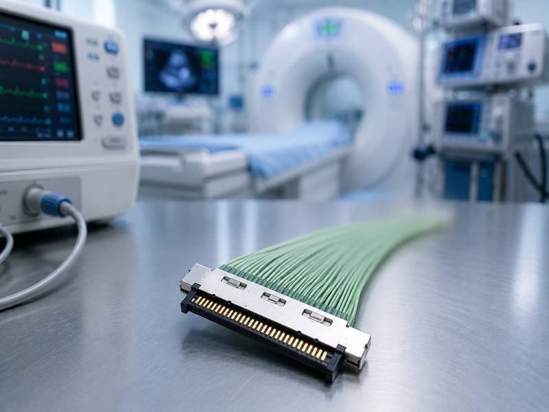
When people think about medical imaging technology, they often focus on the visible components—ultrasound probes, endoscopes, CT scanners, MRI systems, image-processing software, and advanced diagnostic algorithms. Yet some of the most important factors influencing image quality remain hidden deep inside the device. Among these often-overlooked components, micro coax cable assemblies play a surprisingly critical role.
Every medical image begins as an electrical signal. Whether it originates from an ultrasound transducer, a digital imaging sensor, or a detector array inside a CT scanner, that signal must travel through a transmission path before it can be converted into a usable image. If signal degradation occurs during transmission, the resulting image may contain noise, distortion, reduced contrast, or loss of detail. In medical diagnostics, even small reductions in image quality can affect clinical decisions and patient outcomes.
Micro coax cables improve medical imaging quality by enabling high-speed, low-loss signal transmission while maintaining excellent EMI shielding and controlled impedance. Their compact structure allows sensitive imaging data to travel accurately through medical devices, helping reduce image noise, preserve resolution, and support reliable diagnostic performance in ultrasound, endoscopy, CT, MRI, and other advanced imaging systems.
As medical devices become smaller, faster, and more data-intensive, the importance of cable performance continues to grow. Many engineers spend months optimizing software, sensors, and system architecture, only to discover that signal transmission limitations inside the cable assembly are preventing the device from reaching its full performance potential. Understanding how micro coax cables influence imaging quality is no longer a niche engineering topic—it has become a fundamental consideration for modern medical device development. In this article, we will explore why these miniature cable assemblies have such a significant impact on diagnostic imaging and how selecting the right cable solution can contribute to better clinical outcomes.
What Are Micro Coax Cables?
Micro coax cables are miniature coaxial cables designed to transmit high-frequency and high-speed signals in space-constrained medical devices. Their compact size, excellent shielding performance, controlled impedance, and flexibility make them one of the most widely used cable solutions in modern ultrasound, endoscopy, CT, MRI, and other advanced imaging systems.

How Is a Micro Coax Cable Constructed?
Many people are surprised when they first see a micro coax cable. Compared with traditional coaxial cables, it looks incredibly small—sometimes with an outer diameter of less than 0.5 mm. Despite its size, the cable still contains the same fundamental elements required for reliable signal transmission.
At the center is a conductor that carries electrical signals generated by imaging sensors or transducers. Around the conductor sits a precision insulation layer that controls the distance between the conductor and the shield. This spacing is far more important than many non-engineers realize because even tiny dimensional changes can affect impedance stability and signal performance.
Outside the insulation is a metallic shield, typically made from braided copper, aluminum foil, or a combination of both. The shield acts as a protective barrier against electromagnetic interference. Finally, an outer jacket protects the cable from bending, abrasion, chemicals, and mechanical stress.
Although the structure sounds simple, manufacturing a reliable medical-grade micro coax cable is not simple at all. A difference of only a few hundredths of a millimeter in insulation thickness can change electrical characteristics. This is one reason why medical device manufacturers usually require detailed specifications showing dimensions, materials, shielding structure, impedance values, voltage ratings, temperature ranges, and durability performance before approving a cable assembly for production.
When customers contact Sino-Conn about a new medical project, one of the first documents they request is usually a cable specification sheet. Engineers want to verify not only electrical performance but also physical dimensions, flexibility, bend radius, shielding effectiveness, and connector compatibility before moving forward with prototype validation.
How Are Micro Coax Cables Different from Standard Coaxial Cables?
The most obvious difference is size, but in practice size is often the least important reason why medical device manufacturers choose micro coax solutions.
The real advantage is signal density.
A standard coaxial cable may perform perfectly in a laboratory environment, but it quickly becomes impractical when dozens or even hundreds of signal channels must be routed through a compact medical device. Imagine trying to fit 64 or 128 individual standard coaxial cables inside a handheld ultrasound probe. The device would become too large, too heavy, and too uncomfortable for clinical use.
Micro coax cables solve this problem by allowing a large number of signal channels to occupy a much smaller space. This allows engineers to design thinner probes, smaller endoscopes, lighter handheld devices, and more compact imaging systems without sacrificing signal quality.
The comparison below illustrates some of the practical differences.
| Feature | Micro Coax Cable | Standard Coax Cable |
|---|---|---|
| Typical OD | 0.3–1.5 mm | 2–10 mm |
| Flexibility | Excellent | Moderate |
| Signal Density | Very High | Low |
| Medical Imaging Usage | Extensive | Limited |
| Weight | Very Light | Heavier |
| Miniaturized Devices | Ideal | Often Impractical |
Another difference often overlooked is flexibility. Medical devices are rarely stationary. Ultrasound probes are constantly moved. Endoscopes travel through narrow pathways. Robotic surgical systems perform repeated movements throughout procedures.
The cable must survive these motions while continuing to transmit signals accurately. Micro coax cables are specifically designed for these demanding environments and often outperform larger cable constructions when flexibility and durability are equally important.
Which Medical Devices Use Micro Coax Cables?
If you walk through a modern hospital, there is a good chance you will be within a few meters of a device that contains micro coax cable assemblies.
Ultrasound systems are perhaps the most familiar example. Depending on the probe design, dozens or even hundreds of miniature coaxial conductors may be integrated into a single probe cable. These conductors carry signals from transducer elements back to imaging electronics, where they are processed into the images physicians use every day.
Endoscopic imaging systems are another major application. As healthcare providers continue demanding smaller and less invasive procedures, endoscope manufacturers face constant pressure to reduce device diameter while increasing image resolution. This combination creates an ideal environment for micro coax technology.
CT and MRI systems also rely on high-performance signal transmission, although the cable assemblies are often hidden within larger equipment structures. Detector arrays, imaging modules, and communication systems frequently require compact shielded cable solutions capable of handling large amounts of data with minimal signal degradation.
Recent trends in robotic-assisted surgery have created additional demand. Surgical robots rely on cameras, sensors, and precision control systems operating simultaneously. All of these systems require reliable signal transmission in confined spaces. Micro coax assemblies provide a practical solution for managing these requirements.
The table below shows several common applications.
| Medical Device | Typical Use of Micro Coax Cable |
|---|---|
| Ultrasound Probe | Transducer signal transmission |
| Endoscope | HD video transmission |
| CT Scanner | Detector data communication |
| MRI System | RF signal transmission |
| Digital X-Ray | Image sensor connection |
| Surgical Robot | Camera and sensor communication |
| Portable Imaging Device | Compact signal routing |

One interesting trend that many engineers have noticed over the last five years is that cable requirements are becoming more demanding even when devices appear similar externally. Higher image resolutions, AI-assisted diagnostics, real-time image processing, and cloud-connected medical systems all generate more data than previous generations of equipment.
As a result, cable assemblies are no longer viewed as simple accessories. More manufacturers now treat them as critical performance components that directly influence image quality, reliability, and product lifespan.
This is also why many OEM manufacturers involve cable suppliers much earlier in the development process than they did in the past. Instead of waiting until the final design stage, engineering teams increasingly discuss cable routing, connector selection, shielding requirements, and custom assembly options during the early phases of product development. Doing so often reduces redesign costs and shortens the path from prototype to production.
How Do Micro Coax Cables Improve Image Quality?
Micro coax cables improve image quality by preserving weak imaging signals throughout the transmission path. Their combination of controlled impedance, low signal loss, high shielding effectiveness, and compact design helps reduce noise, prevent signal distortion, and maintain image accuracy. In modern ultrasound, endoscopy, CT, and MRI systems, cable performance often has a direct impact on image clarity, diagnostic confidence, and overall device reliability.
How Does Signal Integrity Affect Medical Imaging?
Most imaging problems do not start at the sensor.
This is one of the first lessons many medical device engineers learn during product development.
A sensor may generate an excellent signal. The imaging processor may be capable of handling enormous amounts of data. The software algorithms may be highly optimized. Yet the final image can still appear noisy, unstable, or less detailed than expected.
The reason is simple. The signal must travel from the source to the processor, and every millimeter of that journey matters.
Medical imaging signals are often surprisingly weak. Ultrasound transducers, imaging sensors, and detector arrays generate electrical signals that must be transferred without introducing distortion. During transmission, signals can encounter attenuation, reflections, crosstalk, impedance discontinuities, and external interference. Each of these issues reduces the quality of information reaching the imaging system.
What makes this challenge even more difficult is that modern imaging equipment generates far more data than previous generations. A decade ago, many systems operated comfortably at lower resolutions and lower transmission speeds. Today, manufacturers are integrating HD, 4K, and even higher-resolution imaging technologies into increasingly compact devices.
The margin for transmission errors continues shrinking.
This is where micro coax cables become valuable. Their structure is specifically designed to maintain consistent electrical characteristics across the entire cable assembly. Stable conductor geometry, controlled dielectric spacing, and precision shielding help preserve signal integrity from one end of the system to the other.
Engineers working on ultrasound probes often discover that signal quality improvements achieved through cable optimization can be just as significant as changes made to the transducer itself. That is why many medical OEMs now treat cable assemblies as performance-critical components rather than simple interconnects.
Why Is EMI Shielding Important in Medical Environments?
Hospitals are not electrically quiet places.
A modern imaging system may operate near wireless communication equipment, patient monitoring devices, infusion pumps, surgical systems, networking infrastructure, power supplies, and numerous other electronic devices. Every one of these systems generates electromagnetic energy.

Most people never notice this invisible activity. Sensitive imaging electronics do.
Electromagnetic interference, commonly referred to as EMI, is one of the most common causes of image degradation in electronic systems. When unwanted electromagnetic energy enters a signal path, the result may appear as increased image noise, unstable measurements, reduced contrast, or intermittent imaging artifacts.
The problem becomes more severe as imaging resolution increases.
Higher-resolution systems capture more detail, but they also become more sensitive to interference. A small amount of noise that may have gone unnoticed in older equipment can become a noticeable problem in modern imaging systems.
Micro coax cables help solve this challenge through their shielding structure. Depending on application requirements, manufacturers may use braided shielding, foil shielding, or hybrid shielding systems that combine multiple layers of protection.
The effectiveness of shielding is often measured in decibels (dB). High-quality shielded assemblies may provide shielding effectiveness exceeding 80 dB across specific frequency ranges, dramatically reducing susceptibility to external interference.
When customers approach Sino-Conn regarding medical imaging projects, shielding requirements are often one of the first technical discussions. Many engineers initially focus on connector selection or cable diameter, only to discover later that shielding performance has a greater influence on image quality than expected.
This becomes especially important in portable medical devices where internal electronic components are densely packed into compact housings. Proper shielding design helps ensure that signals remain clean even when operating in challenging electromagnetic environments.
How Does Controlled Impedance Reduce Image Distortion?
Impedance is one of those engineering terms that sounds complicated until a real-world problem appears.
Then it becomes impossible to ignore.
Imagine speaking through a long tunnel. If the tunnel changes shape unexpectedly, your voice may echo, distort, or partially reflect back toward you. Electrical signals behave similarly when they encounter impedance mismatches.
Medical imaging systems often operate at frequencies where signal reflections become a serious concern. When impedance changes unexpectedly along a transmission path, part of the signal can reflect backward. These reflections may cause timing errors, distort waveform shapes, and reduce overall signal quality.
The consequences are not always obvious.
Sometimes image degradation appears as reduced sharpness. In other situations, engineers observe inconsistent performance between units that appear mechanically identical. Troubleshooting these issues can consume weeks of development time.
Micro coax cables are manufactured with tightly controlled dimensions specifically to minimize impedance variation. Consistent conductor size, insulation thickness, and shielding geometry help maintain predictable electrical performance throughout the cable length.
The table below illustrates typical impedance values found in medical imaging applications.
| Application | Typical Impedance |
|---|---|
| RF Systems | 50Ω |
| Video Transmission | 75Ω |
| Specialized Medical Systems | Custom Controlled Values |
What matters most is not simply achieving the correct impedance value but maintaining it consistently across the entire assembly.
This is one reason medical device manufacturers often request detailed impedance specifications and validation reports during supplier qualification. Reliable impedance control helps reduce design risk and contributes directly to better imaging performance.
How Do Low-Loss Materials Preserve Image Accuracy?
Signal loss is unavoidable.
The goal is not to eliminate it entirely but to reduce it to a level where imaging performance remains unaffected.
Every cable introduces some degree of attenuation as signals travel from one point to another. The amount of loss depends on several factors, including conductor quality, insulation materials, operating frequency, cable length, and manufacturing consistency.
As imaging systems generate larger amounts of data, controlling signal loss becomes increasingly important.
Higher-frequency signals are generally more vulnerable to attenuation. If excessive loss occurs, image detail may be reduced before the signal even reaches the processing electronics.
Material selection therefore plays a much larger role than many buyers initially realize.
For example, medical-grade FEP and ETFE materials are frequently selected because they offer excellent dielectric properties, stable electrical performance, and strong environmental resistance. Silicone and TPU materials may be chosen when flexibility becomes the primary concern.
Choosing the wrong material may not immediately cause system failure. Instead, the effects often appear gradually through reduced signal quality, inconsistent performance, or shorter service life.
This is why experienced engineering teams evaluate cable materials based on the complete application environment rather than focusing solely on cost.
A slightly lower-cost material may appear attractive during procurement, but if it contributes to signal degradation or premature replacement, the long-term cost can become significantly higher.
Many OEM customers working with Sino-Conn request material recommendations early in the development cycle for exactly this reason. The best cable solution is rarely determined by a single specification. It is usually the result of balancing signal performance, flexibility, durability, sterilization requirements, environmental exposure, and manufacturing cost.
Why Are Modern Medical Devices Becoming More Dependent on High-Performance Cable Assemblies?
Medical imaging equipment is generating more data than ever before.
A modern ultrasound platform may process significantly more information than systems released only a few years ago. Endoscopy manufacturers are rapidly adopting higher-resolution cameras. AI-assisted imaging platforms require larger datasets to support real-time analysis. Portable imaging devices continue shrinking while simultaneously increasing functionality.
These trends create a common challenge.
More data must move through less space.
The cable assembly has become a critical part of that equation.
In many projects, engineers initially focus on processors, sensors, and software because these components receive the most attention. Later, during system integration, they discover that signal transmission has become the limiting factor.
This shift explains why cable discussions are now occurring much earlier in the development process than they did in the past. Manufacturers increasingly involve cable suppliers during the design stage rather than treating the cable as a component selected after everything else has been finalized.
The result is better system optimization, faster development cycles, and fewer surprises during validation testing.
Ultimately, image quality is not determined by a single component. It is the result of an entire signal chain working together. Micro coax cable assemblies occupy only a small physical space within that chain, but their influence on imaging performance is often far greater than their size would suggest. For many modern medical devices, better images begin with better signal transmission.
Why Are They Used in Ultrasound, Endoscopy, CT, and MRI Systems?
Micro coax cables are widely used in ultrasound, endoscopy, CT, and MRI systems because they can transmit large amounts of sensitive imaging data through extremely limited space while maintaining signal accuracy. Their combination of small size, excellent flexibility, stable impedance, and strong EMI shielding makes them one of the most practical cable solutions for modern medical imaging equipment.
Why Do Ultrasound Systems Require High-Flex Cables?
If you ask medical device engineers where micro coax cables are used most frequently, many will immediately point to ultrasound probes.
The reason becomes obvious once you understand how an ultrasound probe works.
Inside a typical probe, dozens, and sometimes more than a hundred, piezoelectric transducer elements generate electrical signals that must be transmitted back to the imaging system. Every signal contributes to the final image displayed on the screen. The challenge is that all of these signals need to travel through a cable that remains comfortable for physicians to hold and maneuver during examinations.

Modern ultrasound probes are expected to be lightweight, ergonomic, and durable. A bulky cable can make a probe uncomfortable to use, especially during lengthy procedures. At the same time, reducing cable size cannot come at the expense of image quality.
This is where micro coax technology becomes particularly valuable.
A high-end ultrasound probe may contain dozens or even hundreds of miniature coaxial conductors packed into a single flexible cable assembly. The cable must bend repeatedly without affecting signal performance. In busy hospitals, a probe may be used dozens of times per day and experience millions of bending cycles over its lifetime.
Many OEM manufacturers focus heavily on transducer performance, but experienced ultrasound engineers understand that the cable assembly can significantly influence image quality as well. A poorly designed cable may introduce signal loss, increased noise, or inconsistent performance long before the transducer itself reaches its limits.
Another factor often overlooked is probe weight. Reducing cable diameter by only a few millimeters can noticeably improve handling comfort for sonographers who perform hundreds of examinations every month. Better ergonomics not only improve the user experience but can also reduce operator fatigue during long scanning sessions.
This is one reason many ultrasound manufacturers now involve cable suppliers much earlier in development projects. Instead of selecting a cable after completing the probe design, they evaluate routing requirements, shielding performance, flexibility, and cable life expectancy during the early engineering stages.
How Do Endoscopy Systems Benefit from Micro Coax Designs?
Few medical devices demonstrate the value of miniaturization better than modern endoscopes.
Over the last decade, manufacturers have continuously reduced endoscope diameters while simultaneously increasing image resolution. Physicians want better image quality. Patients want less invasive procedures. Achieving both goals requires remarkable engineering compromises.
The internal space available inside an endoscope is extremely limited. Designers must accommodate cameras, LEDs, sensors, control wires, working channels, and signal transmission systems within a very small housing. Every fraction of a millimeter matters.
A traditional cable solution simply occupies too much space.
Micro coax cables allow engineers to transmit high-definition video signals through extremely narrow pathways without sacrificing signal quality. This capability becomes increasingly important as endoscopic systems move toward HD, Full HD, 4K, and even higher imaging standards.
Video quality expectations have changed dramatically. Ten years ago, standard-definition imaging was acceptable for many procedures. Today, physicians expect sharp images capable of revealing small anatomical details. The amount of data transmitted through the cable has increased significantly, while the available space has often decreased.
This creates a unique engineering challenge.
More data.
Less space.
Higher reliability requirements.
Micro coax assemblies solve this challenge by providing excellent signal performance within a compact package. Their controlled impedance characteristics help maintain video signal quality, while their shielding structures reduce interference that could degrade image clarity.
Customers developing endoscopic systems often approach suppliers with requirements that seem contradictory at first. They want smaller cable diameters, higher image quality, improved flexibility, longer service life, and faster development schedules. Meeting all these requirements simultaneously requires careful balancing of conductor size, shielding structure, insulation materials, and connector selection.
Projects like these are rarely solved by simply selecting a standard cable from a catalog. Custom cable assemblies frequently become necessary to achieve the desired performance.
Why Is Signal Stability Critical for CT Imaging?
Computed tomography systems generate enormous amounts of imaging data during every scan.
Although most people think of CT scanners as large machines, many of the engineering challenges inside the system involve transmitting highly sensitive signals with exceptional accuracy. Detector arrays collect vast amounts of information that must be transferred quickly and reliably to processing electronics.
A modern CT scanner may contain thousands of detector channels working simultaneously. Every detector contributes information used to reconstruct detailed cross-sectional images of the human body. Any disruption to signal transmission can potentially affect image reconstruction accuracy.
The challenge becomes greater as CT technology advances.
Higher-resolution imaging requires more detector elements. Faster scanning speeds generate larger amounts of data. Advanced reconstruction algorithms depend on increasingly accurate information.
The cable assembly sits directly in the middle of this process.
Unlike some electronic systems where minor transmission errors may go unnoticed, CT imaging demands consistency. Small signal deviations can sometimes contribute to image artifacts, reduced contrast resolution, or inconsistencies between scans.
This is why engineers often focus heavily on impedance stability, shielding effectiveness, and low-loss signal transmission when selecting cable assemblies for CT-related applications.
The table below illustrates some of the performance priorities commonly encountered in different imaging systems.
| Imaging System | Primary Cable Requirement |
|---|---|
| Ultrasound | Flexibility + Signal Integrity |
| Endoscopy | Miniaturization + Video Quality |
| CT Scanner | High-Speed Data Stability |
| MRI System | EMI Protection + RF Performance |
One interesting trend is that newer CT systems continue increasing data throughput requirements. The cable assemblies used inside these systems must support growing amounts of information without becoming a performance bottleneck. What worked successfully in previous generations may not be sufficient for future platforms.
How Do MRI Systems Minimize Electromagnetic Interference?
MRI environments create some of the most demanding signal transmission conditions found anywhere in healthcare.
Unlike ultrasound or endoscopy systems, MRI technology relies heavily on powerful magnetic fields and highly sensitive radio-frequency signals. These systems are designed to detect subtle changes within the human body, which means they are also extremely sensitive to unwanted interference.
The challenge is not simply transmitting signals.
The challenge is transmitting signals accurately while surrounded by one of the most complex electromagnetic environments found in modern medicine.
Even small amounts of electromagnetic noise can affect signal quality. This makes shielding performance particularly important. Every component involved in signal transmission must be carefully designed to minimize interference and maintain predictable electrical behavior.
Micro coax cables help achieve this through a combination of shielding effectiveness, impedance stability, and compact routing capabilities. Their shield structures help prevent external electromagnetic energy from affecting sensitive signal paths. At the same time, they help contain transmitted signals, reducing the risk of unwanted emissions affecting nearby electronics.
Many engineers working on MRI-related systems pay close attention to shielding specifications because shielding effectiveness can directly influence overall system performance. It is not unusual for engineering teams to request detailed shielding data, impedance reports, and material specifications before approving a cable design.
Another practical benefit is routing flexibility.
MRI systems contain numerous internal subsystems packed into complex mechanical structures. Engineers often need to route cables through confined spaces without compromising performance. The small size of micro coax assemblies makes this task significantly easier compared with larger cable constructions.
As MRI technology continues evolving toward higher field strengths and more advanced imaging techniques, the demands placed on cable assemblies continue increasing. Future systems will likely require even greater attention to signal stability, shielding effectiveness, and manufacturing precision.
Why Are Medical Imaging Manufacturers Increasingly Choosing Custom Cable Assemblies?
A common misconception is that medical device manufacturers simply purchase standard cable assemblies and install them into their products.
The reality is very different.
Most advanced imaging systems have unique mechanical layouts, connector requirements, signal architectures, and regulatory considerations. Off-the-shelf cables often create compromises that limit performance or complicate assembly.
This is why custom cable assemblies have become increasingly common across the medical imaging industry.
A custom solution allows engineers to optimize cable length, routing paths, shielding structures, connector combinations, pin assignments, and material selections for the specific requirements of the device. The result is often improved reliability, easier assembly, reduced weight, and better overall system performance.
Many customers who contact Sino-Conn are surprised to learn how much can be customized. Some initially request only a cable replacement, but after reviewing their application requirements, they discover opportunities to improve flexibility, reduce cable diameter, simplify installation, or shorten lead times.
Another advantage is development speed.
Medical device projects rarely move in a straight line. Specifications change. Connectors become unavailable. Mechanical layouts evolve. Working with a manufacturer capable of providing engineering drawings, prototype support, and customized cable solutions often helps development teams respond more quickly to these changes.
Ultimately, the reason micro coax cables are used so extensively in ultrasound, endoscopy, CT, and MRI systems is not simply because they are small.
They solve a combination of problems that modern medical device manufacturers face every day: transmitting more data, through less space, with higher reliability, while supporting increasingly demanding image quality requirements. As imaging technology continues advancing, their importance is only likely to grow.
Which Cable Specifications Affect Imaging Performance?
Several cable specifications directly influence medical imaging performance, including impedance, shielding effectiveness, conductor quality, dielectric materials, cable diameter, flexibility, bend radius, and connector design. While many buyers initially focus on connector models or cable length, imaging quality is often affected more by the electrical and mechanical characteristics hidden inside the cable itself.
Which Impedance Values Are Common in Medical Imaging?
When engineers begin troubleshooting image quality issues, impedance is often one of the first specifications they review.
The reason is straightforward. Medical imaging systems are designed around predictable signal behavior. If the cable impedance differs from the impedance expected by the imaging electronics, part of the signal can be reflected back toward its source. Those reflections may not always cause obvious failures, but they can gradually reduce signal quality and make image processing more difficult.
Many buyers encounter impedance specifications on drawings without fully understanding their significance. They see values such as 50Ω or 75Ω and assume they are simply numbers required by the design. In reality, those values are directly related to how efficiently signals travel through the cable assembly.
The most common impedance values used in imaging systems are shown below:
| Application | Typical Impedance |
|---|---|
| RF Signal Transmission | 50Ω |
| Video Signal Transmission | 75Ω |
| Custom Medical Systems | Application Specific |
What matters is not only achieving the correct impedance value but maintaining that value consistently across the entire cable assembly. A cable that measures 50Ω in one section and 55Ω in another may still create signal reflections that affect performance.
This is why experienced medical device manufacturers frequently request impedance-controlled cable assemblies instead of generic cable products.
One interesting trend in recent years is that many imaging systems are becoming more sensitive to impedance variation as data rates increase. A design that functioned perfectly five years ago may require much tighter impedance control today because the amount of data being transmitted has increased dramatically.
For OEM customers developing new imaging platforms, impedance verification has become a standard part of qualification testing rather than an optional step.
How Does Shield Coverage Influence Signal Quality?
Shielding is one of those specifications that often receives attention only after a problem appears.
A customer may spend months optimizing sensors, processors, and software. The prototype performs well in the lab. Then the system is tested in a hospital environment and unexpected image noise appears. The root cause frequently turns out to be inadequate shielding.
Medical facilities contain an enormous amount of electronic equipment operating simultaneously. Wireless networks, patient monitoring systems, infusion pumps, surgical tools, communication devices, and imaging equipment all generate electromagnetic energy.
Every imaging signal must travel through this environment without becoming contaminated by unwanted interference.
The effectiveness of shielding depends on several factors, including shield type, coverage percentage, construction quality, and grounding design.
| Shield Type | Typical Advantage |
|---|---|
| Braided Shield | Flexibility and mechanical durability |
| Foil Shield | High-frequency EMI protection |
| Double Shield | Enhanced protection |
| Hybrid Shield | Broad-spectrum EMI control |
A common misconception is that all shielded cables provide the same level of protection. In reality, shielding performance can vary dramatically between different cable constructions.
For example, a cable with 95% braid coverage may provide significantly better protection than one with lower coverage. Likewise, combining foil and braid shielding often delivers better results than relying on either method alone.
Customers developing ultrasound systems frequently ask about shielding effectiveness because ultrasound signals are relatively weak compared to many industrial applications. Even small amounts of interference can affect image consistency.
This is one reason Sino-Conn often discusses shielding requirements before discussing price. A slightly lower-cost cable may initially appear attractive, but if shielding performance is insufficient, the resulting image quality issues can become far more expensive than the original cost savings.
What Role Does Conductor Quality Play in Signal Transmission?
Most people focus on the outside of a cable.
Engineers tend to focus on what is hidden inside.
The conductor is the pathway through which every imaging signal travels. If conductor quality is poor, the entire signal chain can suffer regardless of how advanced the imaging sensor may be.
Several conductor characteristics influence performance:
- Material purity
- Conductor diameter
- Strand structure
- Resistance consistency
- Mechanical durability
Higher-quality conductors generally provide lower resistance and more predictable signal behavior. This becomes particularly important in imaging applications where large amounts of data must be transmitted accurately and repeatedly.
Conductor construction also influences flexibility. A solid conductor may provide certain electrical advantages in specific applications, but stranded conductors are often preferred in medical cable assemblies because they tolerate repeated movement more effectively.
Ultrasound probes provide a good example. These devices experience constant motion throughout daily use. Conductors must survive millions of bending cycles without fracture or performance degradation.
Customers sometimes ask why two seemingly identical cable assemblies have significantly different prices. The answer often lies inside the cable. Differences in conductor quality, material selection, manufacturing tolerances, and testing standards may not be visible externally, but they can have a major impact on long-term performance.
Why Do Dielectric Materials Matter More Than Many Buyers Realize?
When discussing cable specifications, insulation materials rarely receive the same attention as connectors or shielding.
That is understandable because dielectric materials are not visible once the cable assembly is completed. However, they play a major role in determining electrical performance.
The dielectric layer separates the conductor from the shield while helping maintain impedance stability. Different materials influence signal transmission in different ways.
Some materials prioritize electrical performance. Others prioritize flexibility, temperature resistance, chemical resistance, or sterilization compatibility.
The table below summarizes several commonly used materials in medical cable applications.
| Material | Typical Benefits |
|---|---|
| FEP | Excellent dielectric properties and chemical resistance |
| ETFE | High temperature durability |
| TPU | Flexibility and abrasion resistance |
| Silicone | Exceptional flexibility |
| PE | Cost-effective electrical performance |
Choosing the correct material depends heavily on the application environment.
A cable used inside a portable ultrasound system may prioritize flexibility and durability. A cable located inside a sterilizable medical device may require materials capable of surviving repeated cleaning cycles. MRI-related applications may emphasize electrical stability under demanding conditions.
This is why experienced suppliers ask detailed questions about operating environments before recommending materials. A specification that performs well in one application may not be the best choice for another.
How Important Are Cable Diameter and Flexibility?
Medical device engineers often face conflicting requirements.
They want smaller devices.
They want more functionality.
They want better image quality.
And they want longer service life.
Unfortunately, achieving all four simultaneously is rarely simple.
Cable diameter plays a significant role in device design because it directly affects routing flexibility, weight, and available internal space. A reduction of even one or two millimeters can make a substantial difference in handheld devices.
This is particularly true for ultrasound probes and endoscopic systems where space is limited and user comfort matters.
However, reducing diameter introduces new challenges.
Smaller conductors may increase resistance. Tighter constructions can complicate shielding design. Maintaining flexibility while preserving signal quality requires careful engineering.
This balancing act explains why custom cable assemblies have become increasingly popular in medical imaging projects.
Rather than selecting a standard cable and forcing the device design to accommodate it, manufacturers often develop cable assemblies specifically optimized for the intended application.
Customers working with Sino-Conn frequently request reduced-diameter solutions during redesign projects. In many cases, the objective is not simply reducing size. The goal is improving ergonomics, reducing weight, simplifying routing, or enabling a more compact product design.
Why Do Connectors Influence Imaging Performance More Than Expected?
When image quality problems occur, the connector is often overlooked.
Engineers may focus on cable specifications while assuming the connector is merely a mechanical interface. In reality, connectors are critical components of the signal transmission path.
Every connector introduces potential discontinuities.
Poor connector selection can lead to:
- Signal reflections
- Increased insertion loss
- Reduced shielding effectiveness
- Mechanical reliability issues
- Intermittent performance problems
This becomes particularly important in high-speed imaging systems where signal integrity requirements are increasingly strict.
Connector selection involves balancing several factors simultaneously:
| Consideration | Importance |
|---|---|
| Signal Performance | Critical |
| Mechanical Durability | Critical |
| Availability | Important |
| Cost | Important |
| Size Constraints | Critical |
| Assembly Complexity | Moderate |
Many medical device manufacturers now evaluate connectors as part of the overall signal system rather than treating them as independent components.
Customers frequently approach Sino-Conn with connector part numbers and ask whether original or compatible alternatives can be used. In many cases, both options are technically viable. The best choice depends on availability, lead time, budget, regulatory requirements, and long-term supply considerations.
What matters most is ensuring that connector performance supports the overall imaging requirements of the system.
Which Specifications Do Medical Device Manufacturers Prioritize Most?
One of the biggest misconceptions in medical device development is that there is a single “most important” cable specification. In reality, the answer depends largely on where someone sits within the organization. An R&D engineer, a purchasing manager, a quality specialist, and a product manager may all evaluate the same cable assembly differently because each person is responsible for a different part of the project’s success.
For engineering teams, image quality is usually the starting point. Their primary concern is whether the cable can transmit signals accurately without introducing noise, attenuation, impedance instability, or electromagnetic interference. A medical imaging system is only as good as the information reaching the processor. Even the most advanced ultrasound transducer or imaging sensor can struggle to deliver its full potential if signal quality is compromised somewhere along the transmission path. As imaging resolutions continue increasing and data rates continue growing, engineers are paying closer attention to shielding effectiveness, impedance control, insertion loss, and long-term signal stability than ever before.
Mechanical performance is another area that receives significant attention during development. Medical devices are rarely used in ideal laboratory conditions. Ultrasound probes are bent continuously throughout the day. Endoscopic systems are inserted, withdrawn, cleaned, and sterilized repeatedly. Portable imaging equipment may be transported between departments or even different healthcare facilities. Because of this, engineers frequently evaluate flexibility, bend radius, tensile strength, and cable life expectancy alongside electrical performance. A cable that delivers excellent signal quality but fails after a few months of daily use is not considered a successful solution.
The priorities often shift when a project moves from engineering validation into commercial production. At that stage, procurement teams begin focusing on issues such as supply chain stability, lead time, component availability, and overall project cost. A cable assembly may perform exceptionally well during prototype testing, but if critical components require six months of lead time or are difficult to source consistently, production schedules can quickly become unpredictable. Medical device manufacturers cannot afford supply interruptions, particularly when products are already approved and actively used in clinical environments.
Quality and regulatory teams introduce another layer of evaluation. They want to know whether materials are traceable, whether manufacturing processes are repeatable, and whether supporting documentation is available. Certifications such as UL, RoHS, REACH, ISO-related compliance documentation, PFAS declarations, COC, and COO records often become part of the qualification process. In many cases, documentation requirements are nearly as important as the physical product itself because they support regulatory reviews and customer audits.
What makes successful medical imaging projects different is that they balance all of these priorities rather than focusing exclusively on one. The best cable assembly is rarely the cheapest option available, but it is also not automatically the most expensive. The ideal solution is usually the one that delivers stable imaging performance, survives years of daily operation, supports production requirements, and remains available throughout the product lifecycle.
This is one reason many OEM manufacturers involve cable suppliers much earlier in the design process than they did in the past. Rather than waiting until a design is complete, engineering teams increasingly discuss connector options, shielding structures, cable materials, routing constraints, and manufacturing considerations during the development stage. These conversations often reveal opportunities to improve reliability, reduce assembly complexity, shorten lead times, or lower long-term costs before the product reaches production.
From our experience supporting medical imaging projects, the most successful programs typically begin with technical discussions rather than price discussions. Once signal requirements, environmental conditions, mechanical constraints, and compliance requirements are clearly understood, it becomes much easier to identify the right cable solution. When engineering performance, manufacturing practicality, and supply chain considerations are aligned from the beginning, medical device manufacturers are far more likely to achieve both their technical objectives and their commercial goals.
How Do Engineers Choose the Right Medical Micro Coax Cable?
Choosing the right medical micro coax cable requires more than matching a connector or selecting a cable diameter. Engineers must evaluate signal requirements, impedance control, shielding performance, flexibility, durability, materials, connector compatibility, and long-term reliability. The best cable is the one that supports both imaging performance and the practical realities of manufacturing, sterilization, maintenance, and product lifecycle management.
What Information Should Be Included in a Cable Specification?
One of the most common situations in the medical cable industry is surprisingly simple: a customer knows exactly what the device needs to do but does not have a complete cable specification.
Sometimes an engineer sends a part number. Sometimes a purchasing manager sends a photograph. Sometimes a maintenance department sends an old cable and asks whether it can be duplicated. In many cases, the original documentation no longer exists, especially for legacy medical equipment that has been in service for many years.
The challenge is that medical imaging cables are rarely defined by a single parameter. A complete specification usually includes electrical, mechanical, environmental, and regulatory requirements.
A properly documented medical micro coax cable specification often includes:
| Specification Category | Typical Requirements |
|---|---|
| Electrical | Impedance, voltage, current, shielding |
| Mechanical | OD, bend radius, flexibility |
| Materials | Jacket, dielectric, conductor type |
| Connector | Model, orientation, mating requirements |
| Environmental | Temperature, sterilization, chemical resistance |
| Compliance | UL, RoHS, REACH, PFAS declarations |
The reality is that many projects begin without all of this information. This is why experienced cable manufacturers spend significant time asking questions before providing quotations.
For example, a cable that will be installed permanently inside a CT scanner has very different requirements from a cable used on an ultrasound probe that may be bent thousands of times every week. The connector may be identical, but the cable construction could be completely different.
Many customers are surprised when a supplier asks detailed questions about routing space, expected service life, sterilization methods, or bending frequency. Those questions are not slowing down the project. They are usually helping prevent problems that would otherwise appear months later during validation or production.
How Important Are Drawings Before Production?
Many of the most expensive cable-related mistakes occur before production even begins.
A connector is rotated 180 degrees.
A pin assignment is misunderstood.
A cable length is interpreted differently.
A strain relief is positioned incorrectly.
None of these problems are particularly complicated, but all of them can delay a project and create unnecessary costs.
This is why engineering drawings remain one of the most valuable tools in custom cable development.
An approved drawing creates a common technical language between the customer and manufacturer. Instead of relying on assumptions, both sides review the exact dimensions, pin definitions, connector orientations, materials, labels, and assembly requirements before production starts.
Experienced engineers understand the value of this step because finding an error on a drawing is far less expensive than finding the same error after production.
Many OEM customers now treat drawing approval as a formal milestone in their development process. Once the drawing is approved, both parties know exactly what is being manufactured.
This is one area where response speed can significantly affect project timelines. For urgent projects, waiting two weeks for a drawing review is often unacceptable.
Sino-Conn regularly supports customers who require rapid engineering feedback. Depending on project complexity, drawings can often be prepared within a few days, and in some straightforward projects preliminary engineering support may be available much faster. This helps engineering teams move forward without unnecessary delays while still maintaining proper design control.
Should You Choose Original or Compatible Connectors?
This question appears in almost every medical cable project at some stage.
Engineers frequently specify original connector brands because they are familiar with their performance and have used them successfully in previous designs. There is certainly value in this approach, particularly when a device has already completed qualification testing or regulatory approvals.
However, real-world manufacturing introduces challenges that engineering drawings do not always reveal.
A connector may suddenly have a long lead time.
Inventory may become limited.
Minimum order quantities may increase.
A new project may require a connector modification that the original manufacturer does not support.
These situations often lead companies to evaluate compatible alternatives.
The discussion should not focus solely on price. Performance, reliability, fit, availability, and long-term supply stability are all important considerations.
Many medical device manufacturers discover that compatible connectors can provide similar functionality while offering greater flexibility for customization and shorter delivery schedules. Others determine that maintaining the original connector remains the best decision for regulatory or qualification reasons.
The right answer depends on the specific application rather than a general rule.
One thing that experienced OEM manufacturers have learned is that connector selection should be evaluated early in development rather than after the design is finalized. Making connector decisions during the early stages often prevents supply chain surprises later in the project.
How Do Engineers Evaluate Cable Reliability Before Product Launch?
Medical imaging systems are expected to operate reliably for years, not weeks.
That expectation creates a challenge during development because engineers must predict how a cable will perform long before the product reaches the market.
The most effective way to evaluate reliability is through testing that simulates real-world conditions.
For an ultrasound probe, this may involve repeated bending tests.
For a portable imaging device, it may involve movement, vibration, and handling simulations.
For hospital equipment, environmental testing may include temperature cycling, humidity exposure, and cleaning chemical compatibility.
The goal is not simply proving that a cable works today. The goal is gaining confidence that it will continue working after years of daily use.
This is particularly important because cable failures are often difficult to diagnose once equipment is deployed. A cable may perform perfectly during initial installation and then gradually develop intermittent issues after thousands of operating cycles.
Many medical OEMs therefore place significant value on validation data, quality records, and manufacturing consistency. Reliable suppliers understand that customers are not only purchasing a cable assembly. They are purchasing confidence that the product will continue performing as expected throughout its intended service life.
What Questions Should Engineers Ask a Cable Supplier?
The quality of a project is often influenced by the quality of the questions asked during the early stages.
Many first-time buyers focus almost entirely on price and delivery. Experienced engineers usually take a broader view.
Instead of asking only “How much does it cost?”, they ask questions such as:
Can the cable maintain controlled impedance?
How effective is the shielding structure?
What materials are used in the construction?
How is long-term reliability verified?
What testing is performed before shipment?
How quickly can engineering changes be implemented?
Can the supplier support future production volumes?
These questions often reveal far more about a supplier’s capabilities than pricing alone.
In medical imaging applications, technical support can be just as valuable as manufacturing capability. A supplier that understands signal integrity, shielding requirements, connector compatibility, and regulatory expectations can help identify potential risks before they become expensive problems.
This is one reason many medical device manufacturers prefer working directly with cable assembly specialists during development. The earlier technical discussions occur, the easier it becomes to optimize performance, simplify manufacturing, and avoid redesigns.
Why Are More Medical Device Manufacturers Involving Cable Suppliers Earlier?
Ten years ago, cable assemblies were often selected near the end of a project.
The device was designed first.
The cable was selected afterward.
That approach is becoming increasingly rare.
Modern imaging systems generate more data, occupy less space, and operate under stricter performance requirements than previous generations. As a result, cable assemblies have become integral parts of the overall system architecture rather than simple accessories.
When cable suppliers become involved early, engineers gain access to practical information about connector availability, routing constraints, shielding options, material selection, manufacturability, and production planning. These discussions frequently identify opportunities for improvement that would be difficult to recognize later.
The benefits are often measurable.
Shorter development cycles.
Fewer redesign iterations.
Better signal performance.
Improved manufacturability.
Reduced supply chain risk.
For medical device manufacturers competing in increasingly demanding markets, these advantages can have a meaningful impact on both project timelines and long-term product success.
Ultimately, selecting the right medical micro coax cable is not about finding a component that meets a specification sheet. It is about finding a solution that supports image quality, reliability, manufacturability, regulatory compliance, and future production requirements simultaneously. The most successful projects are usually the ones where those considerations are addressed together from the very beginning rather than one at a time later in development.
Can Custom Cable Assemblies Improve Device Development?
Quick Answer
Custom cable assemblies often improve medical device development by optimizing signal performance, reducing space constraints, simplifying assembly processes, and supporting unique design requirements that standard products cannot address.
Which Cable Parameters Can Be Customized?
No two medical devices are exactly alike. Even products serving similar functions may have different space constraints, signal requirements, connector preferences, and environmental conditions. This is why custom cable assemblies have become increasingly important throughout the medical device industry.
Customization extends far beyond cable length. Engineers may require specific connector combinations, unique pin assignments, specialized shielding structures, custom materials, low-profile routing solutions, or enhanced flexibility characteristics. In some applications, the goal is to maximize performance. In others, the objective may be reducing assembly complexity or improving long-term reliability.
Common customization options include cable length, connector type, pinout configuration, shielding design, outer jacket material, conductor construction, overmolding features, strain relief design, labeling requirements, and packaging specifications.
The ability to customize these parameters allows manufacturers to optimize cable performance for the exact requirements of the device rather than forcing the device to accommodate a standard cable product. Over time, this flexibility often contributes to improved system performance, easier assembly, and lower overall development costs.
How Can Rapid Prototyping Accelerate Medical Projects?
Speed is becoming increasingly important in medical device development. Competitive markets, evolving regulations, and shorter product life cycles place pressure on engineering teams to move quickly while maintaining quality standards.
Prototype cable assemblies play a critical role during development because they allow engineers to validate designs before committing to full-scale production. Early prototypes help identify potential issues related to routing, connector fit, signal performance, flexibility, and manufacturability.
A slow prototyping process can delay the entire development schedule. Conversely, rapid prototype availability allows teams to test, refine, and improve designs earlier in the development cycle. This accelerated feedback loop often reduces overall project risk and shortens time-to-market.
One reason many OEM manufacturers work closely with specialized cable assembly suppliers is the ability to obtain engineering support and prototype assemblies quickly. Sino-Conn supports this process through rapid drawing preparation, engineering consultation, and flexible sample production capabilities. For urgent projects, expedited sample development can significantly reduce waiting time and help engineering teams maintain project momentum.
How Does Custom Design Solve Space Constraints?
Medical devices continue becoming smaller, lighter, and more portable. While these trends benefit clinicians and patients, they create significant challenges for design engineers. Every millimeter of available space must be used efficiently, and cable assemblies often become one of the most difficult components to integrate.
Standard cable products may perform well electrically but consume excessive space. They may also require larger bend radii or create routing conflicts within compact assemblies. Custom cable designs address these challenges by tailoring dimensions, connector configurations, and routing characteristics to the specific device architecture.
Micro coax cable assemblies are particularly effective in compact systems because they support high-density signal transmission without sacrificing performance. By combining miniature conductors with optimized connector solutions, engineers can reduce cable volume while maintaining reliable signal transmission.
As wearable medical devices, portable imaging systems, and robotic surgical platforms continue evolving, the ability to develop highly customized cable solutions will become increasingly valuable.
What Future Trends Are Driving Medical Micro Coax Cable Development?
Quick Answer
Future medical micro coax cable development is being driven by device miniaturization, higher imaging resolutions, AI-assisted diagnostics, increased data transmission requirements, and the growing demand for flexible, high-performance cable assemblies.
How Is Medical Device Miniaturization Changing Cable Design?
Medical technology has steadily moved toward smaller and more portable systems. Devices that once occupied entire rooms are now available as compact mobile units. Portable ultrasound systems, wearable monitoring devices, and handheld diagnostic tools illustrate this trend clearly.
As devices shrink, cable assemblies must also become smaller without compromising performance. This challenge requires innovations in conductor design, insulation materials, shielding structures, and manufacturing processes. Engineers increasingly seek cable solutions capable of supporting high data rates within extremely compact dimensions.
Miniaturization also increases the importance of mechanical flexibility. Smaller devices often contain tighter routing paths and more complex internal layouts. Cable assemblies must adapt to these constraints while maintaining long-term reliability.
How Will AI-Assisted Diagnostics Increase Data Requirements?
Artificial intelligence is rapidly transforming medical imaging. Advanced algorithms now assist physicians by identifying abnormalities, improving image reconstruction, and enhancing diagnostic workflows. However, AI systems depend heavily on data quality.
Higher-resolution imaging generates larger datasets that must be transmitted quickly and accurately. As imaging systems evolve, cable assemblies must support increasing data volumes without introducing errors or signal degradation.
The relationship between AI and cable technology may not appear obvious at first, but it is becoming increasingly important. Advanced software can only perform effectively when it receives accurate data. Reliable signal transmission therefore remains a foundational requirement for future AI-driven healthcare systems.
What Problems Can Poor Cable Design Cause?
Poor cable design can create far more than simple connectivity issues. In medical imaging systems, it can contribute to image noise, signal loss, intermittent failures, reduced device reliability, increased maintenance costs, longer development cycles, and even delayed product launches. Many imaging performance problems that initially appear to be software or hardware issues can ultimately be traced back to the cable assembly itself.
Why Does Image Noise Increase Over Time?
One of the most frustrating situations for engineers is when a system passes validation testing but begins exhibiting image quality problems several months later.
The imaging sensor has not changed.
The software has not changed.
The imaging algorithm remains the same.
Yet somehow image noise gradually becomes more noticeable.
In many cases, the problem is not the imaging system itself. It is the cable assembly aging under real-world operating conditions.
Medical cables experience stress every day. Ultrasound probes are bent repeatedly. Portable imaging devices are transported between departments. Endoscopy systems undergo cleaning and sterilization cycles. Over time, these mechanical and environmental stresses can affect shielding performance, conductor integrity, and insulation stability.
The degradation is often gradual rather than catastrophic.
Instead of a complete failure, engineers may notice subtle changes such as increased background noise, reduced image contrast, intermittent artifacts, or inconsistent image quality between devices.
This is one reason why experienced medical device manufacturers pay close attention to life-cycle testing. A cable that performs perfectly during initial validation may not deliver the same performance after one million bending cycles.
For ultrasound applications, repeated flexing is particularly demanding. Some probe cables experience thousands of bends every day. If conductor construction, shielding design, or strain relief structures are not optimized correctly, performance degradation can occur much sooner than expected.
The challenge is that these issues often appear long after product launch, making them more expensive to diagnose and correct.
How Can Poor Shielding Affect Diagnostic Accuracy?
Most hospitals contain far more electromagnetic activity than people realize.
Wireless networks, patient monitors, infusion systems, communication equipment, surgical devices, and imaging platforms all operate simultaneously. Every one of these systems generates electromagnetic energy.
A well-designed cable assembly protects imaging signals from this environment.
A poorly designed one does not.
When shielding effectiveness is inadequate, unwanted electromagnetic interference can enter the signal path. The result may be image artifacts, reduced contrast, unstable video transmission, or inconsistent measurements.
The impact is not always dramatic.
In some cases, the image still appears acceptable to the human eye, but subtle diagnostic details become more difficult to distinguish. This becomes increasingly important as imaging systems move toward higher resolutions and more advanced diagnostic applications.
A common misconception is that all shielded cables perform similarly. In reality, shielding performance can vary significantly depending on shield coverage, material quality, grounding design, and manufacturing consistency.
For example, a cable with incomplete shield coverage may perform adequately in a laboratory environment but struggle when installed in a hospital filled with electronic equipment. This is one reason many medical OEMs request shielding specifications and validation data during supplier qualification.
The cost difference between a basic shielded cable and a properly optimized shielded assembly is often small compared to the cost of troubleshooting imaging issues after deployment.
What Causes Signal Loss in Medical Imaging Systems?
Signal loss is one of the most overlooked performance issues during early-stage development.
Many engineers focus heavily on connectors, sensors, and processors because those components appear to have the greatest influence on image quality. The cable is often viewed as a passive component simply transferring information from one point to another.
In reality, every cable introduces some degree of signal attenuation.
The question is whether that attenuation remains within acceptable limits.
Several factors contribute to excessive signal loss:
- Poor conductor quality
- Inconsistent impedance
- Low-quality dielectric materials
- Excessive cable length
- Inadequate connector design
- Manufacturing inconsistencies
As imaging systems generate larger volumes of data, signal loss becomes increasingly important. Higher-frequency signals are generally more sensitive to transmission imperfections. A cable design that performed adequately for older imaging systems may become problematic in next-generation devices.
One situation seen frequently during prototype development involves customers attempting to reduce costs by selecting lower-grade materials. The cable appears identical from the outside, but differences in conductor purity or dielectric quality can significantly affect transmission performance.
The result is often a difficult troubleshooting process. Engineers investigate software, sensors, and electronics before eventually discovering that the cable assembly itself is limiting performance.
How Can Repeated Flexing Damage Internal Conductors?
Many medical cables spend their entire lives moving.
This reality is easy to overlook during design reviews because engineers usually examine cable drawings while the cable is perfectly straight.
Actual operating conditions are very different.
An ultrasound probe cable may be twisted, bent, and repositioned hundreds of times during a single day. Endoscopic systems experience repeated movement throughout procedures. Portable imaging devices are frequently transported and handled by multiple users.
Every movement introduces mechanical stress.
Over time, these stresses accumulate.
Eventually, conductors may begin developing microscopic fractures. Shield structures may lose effectiveness. Insulation layers may experience fatigue. The cable continues functioning, but performance becomes less predictable.
Intermittent failures are particularly challenging because they are difficult to reproduce during troubleshooting.
The system works.
Then it doesn’t.
Then it works again.
Many engineers consider intermittent failures among the most time-consuming problems to diagnose because they often appear random.
This is why cable flexibility and bend-life performance are not secondary specifications in medical applications. They are often critical reliability requirements that directly influence product lifespan.
Manufacturers developing portable or handheld imaging systems increasingly request bend-life validation data before approving cable assemblies for production. They want confidence that the cable will survive years of real-world use rather than simply passing laboratory testing.
Why Do Some Medical Imaging Projects Experience Unexpected Delays?
Not all cable-related problems occur after a product launches.
Some occur much earlier.
A surprising number of medical imaging projects experience development delays because cable requirements are considered too late in the design process.
A connector becomes unavailable.
The cable routing is physically impossible.
The shielding performance is insufficient.
The cable diameter is larger than expected.
The required materials have extended lead times.
These issues may seem minor individually, but together they can significantly delay validation schedules and product launches.
One trend that has become increasingly common is involving cable manufacturers during the early stages of product development rather than after the design is finalized. Early discussions often identify potential risks before they become expensive redesigns.
Many Sino-Conn customers initially contact us for quotations and later discover that engineering support is equally valuable. By reviewing cable routing requirements, connector availability, shielding needs, and manufacturing feasibility early in the process, development teams can often avoid weeks or even months of unnecessary delays.
The earlier potential issues are identified, the easier and less expensive they are to solve.
What Are the Hidden Costs of Cable Failure?
When people calculate cable costs, they usually focus on the purchase price.
The actual cost of failure is often much higher.
Consider a medical imaging device that experiences intermittent signal problems in the field.
The manufacturer may need to:
- Diagnose the issue
- Replace components
- Provide technical support
- Ship replacement assemblies
- Conduct failure analysis
- Manage customer dissatisfaction
- Delay future product shipments
The cost of these activities can quickly exceed the original value of the cable assembly itself.
In regulated industries such as healthcare, the stakes become even higher. Product reliability affects not only operational costs but also customer trust and brand reputation.
This is why many experienced medical device manufacturers view cable assemblies as strategic components rather than commodities.
A slightly higher initial investment in cable quality often reduces long-term costs by improving reliability, reducing maintenance requirements, and minimizing the risk of field failures.
The most successful imaging systems rarely achieve their performance through a single breakthrough component. They achieve it through the consistent performance of hundreds of interconnected parts working together. The cable assembly may occupy only a small portion of the overall system, but when it is poorly designed, the consequences can affect everything from image quality and reliability to development schedules and customer satisfaction.
For medical imaging manufacturers, avoiding these problems is often far less expensive than fixing them later.
How Does Sino-Conn Support Medical Imaging Projects?
Sino-Conn supports medical imaging projects through engineering collaboration, custom cable development, rapid drawing support, prototype manufacturing, flexible connector sourcing, and multi-stage quality control. Rather than simply supplying cable assemblies, the focus is on helping medical device manufacturers reduce development risk, shorten project timelines, and achieve reliable long-term imaging performance.
How Can Sino-Conn Help When Technical Information Is Incomplete?
One of the biggest misconceptions in custom cable manufacturing is that every customer arrives with a complete drawing package and a detailed specification sheet. In reality, that is often not the case.
Many medical imaging projects begin with surprisingly limited information. An engineer may have only a connector model number. A purchasing manager may only have a photograph from an existing system. In some situations, customers send a worn cable assembly removed from equipment that has been operating in hospitals for years and ask whether an equivalent replacement can be developed.
This happens frequently in the medical industry because product lifecycles are often much longer than in consumer electronics. Imaging equipment may remain in service for ten years or more, while original suppliers, part numbers, or documentation may no longer be available.
The first step is therefore not manufacturing. It is understanding the application.
Before discussing pricing or production schedules, engineering teams typically review the available information, identify connectors, evaluate cable structures, and understand how the assembly functions within the imaging system. Questions regarding signal transmission, cable routing, sterilization requirements, bend-life expectations, and environmental conditions are often more important than the initial cable dimensions themselves.
This approach frequently helps customers uncover potential issues that would otherwise remain hidden until later development stages. In many projects, the discussion evolves from “Can you copy this cable?” to “Can we improve the design while maintaining compatibility?” That shift often creates opportunities to reduce cable diameter, improve flexibility, shorten lead times, or increase long-term reliability.
Why Is Engineering Support So Important in Medical Cable Projects?
A medical imaging cable assembly may appear simple when viewed from the outside, but its role inside the device is rarely simple.
Every imaging signal generated by a transducer, sensor, detector, or camera must travel through a transmission path before becoming a usable image. Any weakness in that path can affect overall system performance. Because of this, cable-related decisions are increasingly being made during product development rather than after the device design is completed.
Many customers approach Sino-Conn during the early stages of development because they need practical engineering feedback rather than just manufacturing capacity. They may be evaluating connector options, determining whether a cable can survive repeated flexing, reviewing shielding requirements, or trying to reduce the overall size of an imaging subsystem.
These discussions often have a measurable impact on project timelines. A routing issue discovered during the design stage may take a few hours to solve. The same issue discovered after tooling, validation testing, or pilot production may delay a project by weeks.
The table below illustrates some of the engineering topics most frequently discussed during medical imaging projects.
| Development Area | Typical Customer Concern |
|---|---|
| Signal Integrity | Image quality and data accuracy |
| Shielding Design | EMI protection and noise reduction |
| Connector Selection | Availability and compatibility |
| Cable Routing | Space limitations inside devices |
| Material Selection | Flexibility and environmental resistance |
| Manufacturing | Scalability and production readiness |
The earlier these topics are addressed, the easier it becomes to avoid expensive redesigns later in the project lifecycle.
How Fast Can Drawings, Samples, and Production Be Delivered?
Development schedules rarely move as slowly as engineers would like.
Medical device companies are often balancing multiple pressures simultaneously. Product launch targets, investor expectations, customer commitments, regulatory milestones, and internal development deadlines all influence the pace of a project.
This is one reason response speed has become such an important factor when selecting a cable assembly supplier.
A technically capable supplier that requires several weeks to respond to engineering requests may unintentionally slow an entire development program. On the other hand, rapid engineering feedback allows teams to evaluate ideas, test alternatives, and move projects forward much more efficiently.
The support timeline below reflects the capabilities that many customers rely on during development and production.
| Project Activity | Typical Timeline |
|---|---|
| Initial Technical Review | Same Day |
| Engineering Evaluation | Within 24 Hours |
| Drawing Preparation | 30 Minutes to 3 Days |
| Standard Sample Build | Approximately 2 Weeks |
| Urgent Prototype Samples | 2–3 Days |
| Standard Production Orders | 3–4 Weeks |
| Expedited Production Orders | Approximately 2 Weeks |
Of course, not every project follows the same schedule. A simple adapter cable and a highly customized medical imaging harness require very different levels of engineering effort. What matters most is maintaining realistic expectations and transparent communication throughout the process.
Many OEM customers report that predictable communication often provides just as much value as production speed. Knowing when drawings, samples, or production units will be available allows engineering and procurement teams to plan effectively and avoid unnecessary delays.
How Does Sino-Conn Support Custom Medical Cable Development?
Standard products rarely solve every challenge in medical imaging equipment.
As devices become smaller and imaging performance requirements continue increasing, manufacturers frequently require cable assemblies that cannot be purchased directly from a catalog.
A modern ultrasound probe may require an extremely flexible cable capable of surviving millions of bending cycles. An endoscopic imaging system may need high-density signal transmission within an exceptionally limited space. A portable imaging platform may require a lighter cable construction without sacrificing shielding effectiveness.
Each application introduces unique constraints.
This is why customization has become such an important part of medical cable development.
The most common customization requests typically involve cable length, connector combinations, pin assignments, shielding structures, outer jacket materials, cable diameter, and mechanical routing requirements. In many projects, the objective is not simply replicating an existing cable but improving performance while maintaining compatibility with the device architecture.
The ability to customize these parameters often allows engineers to optimize system performance rather than forcing the system to accommodate a standard cable solution. Over time, this can contribute to better image quality, improved reliability, and simplified assembly processes.
How Does Sino-Conn Balance Original and Compatible Connector Solutions?
Connector sourcing has become increasingly complex in recent years.
Many medical device manufacturers prefer original branded connectors because they have proven performance records and established qualification histories. In some applications, maintaining the original connector remains the most practical choice.
However, real-world supply chains introduce additional considerations. Lead times fluctuate, inventory availability changes, and some projects require greater flexibility than original manufacturers can provide.
Rather than approaching connector selection from a purely cost-driven perspective, most medical OEMs evaluate the broader picture.
| Evaluation Factor | Original Connector | Compatible Connector |
|---|---|---|
| Performance Consistency | Excellent | Application Dependent |
| Lead Time | Often Longer | Often Shorter |
| Availability | Variable | More Flexible |
| Custom Modification | Limited | More Flexible |
| Cost | Higher | Lower |
The best choice depends on project requirements, qualification considerations, production volume, and long-term supply objectives.
Many customers evaluate both options during development to reduce future supply chain risk while maintaining confidence in system performance.
How Does Sino-Conn Ensure Consistent Quality?
Image quality ultimately depends on signal quality, and signal quality depends heavily on manufacturing consistency.
A medical cable assembly may contain dozens of individual manufacturing processes before it reaches the customer. Connector termination, conductor preparation, shielding treatment, soldering quality, overmolding, routing, and final assembly all influence the finished product.
Even small variations can affect performance.
This is why medical device manufacturers often spend considerable time evaluating quality systems before approving a supplier.
Rather than relying exclusively on final inspection, quality verification occurs throughout the manufacturing process.
| Quality Stage | Purpose |
|---|---|
| Incoming Inspection | Verify raw materials and components |
| Process Inspection | Monitor manufacturing consistency |
| Final Inspection | Confirm finished product quality |
| Pre-Shipment Inspection | Verify shipment readiness |
In practical terms, this means assemblies are checked multiple times before leaving the factory. For customers developing medical imaging equipment, this approach helps improve consistency across prototypes, pilot runs, and production orders.
Why Do Many Medical OEMs Continue Working with the Same Supplier?
The answer is rarely based on pricing alone.
By the time a medical imaging product reaches production, manufacturers have often invested significant time validating every critical component within the system. Engineering teams have completed performance testing, quality departments have reviewed compliance documentation, production teams have established assembly procedures, and regulatory personnel have incorporated approved components into qualification records.
Changing suppliers after that point can create additional testing requirements, documentation updates, qualification activities, and potential project risk. Because of this, many OEM manufacturers place significant value on stability and long-term support.
Over time, the relationship often extends beyond manufacturing. Engineering teams may rely on supplier feedback when evaluating new connector options. Procurement departments benefit from predictable lead times and stable communication. Production teams gain confidence from consistent quality and repeatable manufacturing processes.
The companies that remain successful in medical imaging rarely view cable assemblies as simple commodities. They recognize that reliability, engineering support, responsiveness, and manufacturing consistency often have a greater impact on long-term project success than small differences in unit cost.
For this reason, many customers work with Sino-Conn not only as a cable assembly manufacturer but as a long-term engineering partner. Whether the requirement involves a replacement cable, a new imaging platform, a prototype concept, or a fully customized medical harness, the objective remains the same: reduce development risk, improve reliability, and help bring medical imaging products to market faster and more efficiently.
Conclusion
Medical imaging quality depends on far more than sensors, software, and processing power. Behind every clear ultrasound image, high-resolution endoscopic video, CT scan, or MRI result lies a complex signal transmission system responsible for preserving data accuracy from source to processor.
Micro coax cables play a critical role in this process. Their ability to provide excellent signal integrity, effective EMI shielding, controlled impedance, compact routing, and long-term reliability makes them an essential component of modern medical imaging equipment. As healthcare technology continues evolving toward higher resolutions, faster data rates, AI-assisted diagnostics, and smaller device architectures, the importance of high-performance cable assemblies will only continue to grow.
For engineers, OEM manufacturers, and medical device developers, selecting the right cable assembly partner can be just as important as selecting the right imaging technology. The most successful projects often begin with detailed technical discussions, careful specification reviews, and close collaboration between engineering teams and manufacturing specialists.
Whether you have a complete drawing package, a prototype concept, a legacy cable sample, or simply a photo of an existing assembly, the Sino-Conn engineering team can help evaluate your requirements and develop a customized solution tailored to your application.
Need a custom medical micro coax cable assembly for your imaging system? Contact Sino-Conn today for engineering consultation, drawing support, rapid prototyping, and reliable manufacturing solutions designed specifically for modern medical devices.